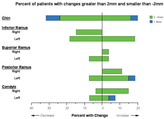
Fig 9.
Percentages of patients with changes (between splint removal and 1-year follow-up) >2 mm and <−2 mm for the 9 anatomic regions of interest. Patients with displacements between −2 and 2 mm are not represented. Note that positive or negative values of displacements represent different directional movements depending on the specific region of interest.

