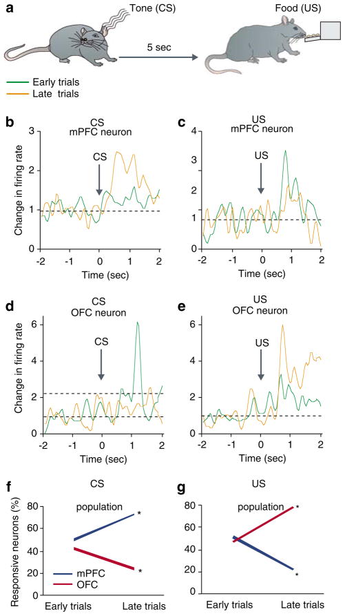
Figure 2.
PFC subregions display differential patterns of plasticity during a classical conditioning paradigm. (a) Rats were trained inside an operant chamber to associate a tone (CS) with food reward (US) delivered 5 s later. Each rat received 18 presentation of CS with an inter-CS interval of 120 s. (b–e) Single neurons recorded from the mPFC or OFC exhibited opposite plastic responses to task relevant events of CS and US. Examples compare the phasic peri-event time histograms (window=±2 s around event, bin=50 ms) of individual neurons during early and late eight trials within the same learning session. Note that the mPFC neuron in (b) amplified its representation of CS, whereas the other mPFC neuron in (d) loses its initial response to US. In contrast, the OFC neuron in (c) diminishes its representation of CS, whereas the OFC neuron in (e) amplifies its response to UCS. Firing rates are normalized to pre-tone baseline period. (f–g) Plasticity in the population response of the two regions assessed by the proportion of neurons with a phasic response. The mPFC and OFC display opposite trajectories; the mPFC enhanced its representation of CS and OFC that of US. *p<0.05 compared to early trials (based on 136 mPFC and 94 OFC neurons).

