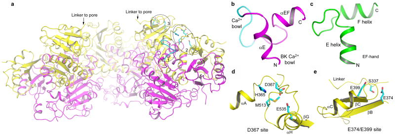
Figure 3.
Ca2+ binding sites in the BK intracellular subunit. a, A side view of the BK gating ring with the top surface facing the membrane. The front subunit is removed for clarity. The circled positions represent the three Ca2+ binding sites on one subunit, and are labeled 1 for the Ca2+ bowl, 2 for the Asp367 site and 3 for the Glu374/Glu399 site. b, αE, the Ca2+ bowl, and αEF form an EF-hand like motif. The structure of the Ca2+ bowl (cyan loop) is disordered in the absence of Ca2+. c, The EF-hand structure of a Ca2+ binding protein (a human Calmodulin molecule with PDB code 1CLL). d and e, Structural details of the Asp367 and E374/E399 Ca2+ binding sites, respectively.

