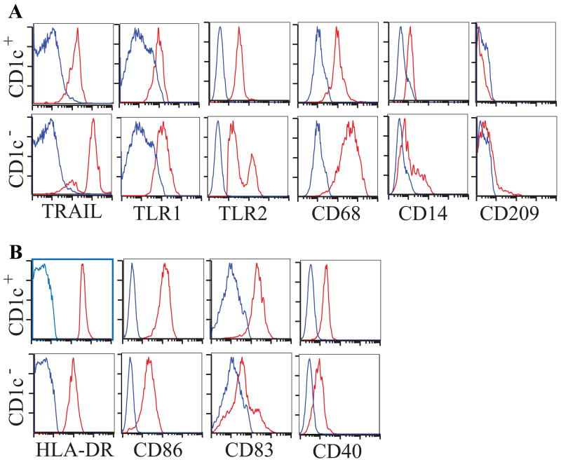
Figure 3. Flow cytometric analysis of psoriatic dermal single cell suspensions.
(A) CD1c- DCs (bottom row) express higher levels of TRAIL, TLR1, TLR2, CD68, CD14 and CD209 compared to CD1c+ DCs (top row), (B) CD1c+ DCs expressed comparatively higher levels of DC maturation antigens HLA-DR, CD86, CD83 and CD40. Red histogram represents antigen expression gated on HLA-DR+CD11c+CD163-CD1c+/- DCs, blue is isotype.

