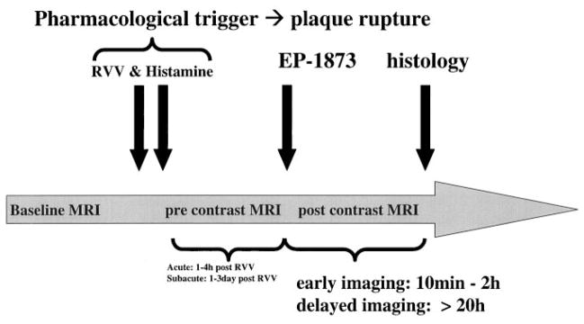
Figure 1.
Time course of in vivo MRI experiment of acute and subacute thrombosis with the use of an atherosclerotic New Zealand White rabbit model. After an 8-week high-cholesterol diet, baseline MRI of the subrenal aorta was performed. Subsequently, plaque rupture was induced with the use of RVV and histamine. The fibrin-binding peptide derivative EP-1873 was then intravenously administered ≈1 hour (imaging of acute thrombosis) or ≈20 hours (imaging of subacute thrombosis) after RVV and histamine. After intravenous injection of EP-1873, MRI was performed at 30 minutes and continued up to ≈20 hours. Subsequently, the animals were killed, and histological analyses were performed.

