
Figure 4.
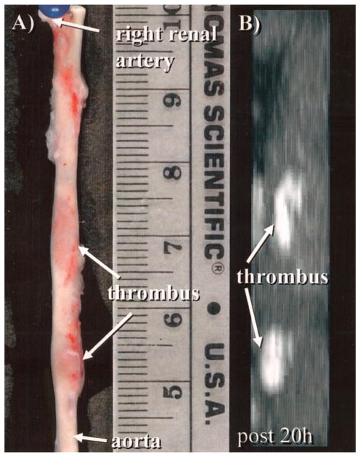
A, Photograph demonstrates 2 well-delineated focal thrombi (arrows) in a harvested subrenal aorta. B, On corresponding targeted MIP, 2 bright thrombi (arrows) can be identified that correspond in both size and location with gross histological image in A.