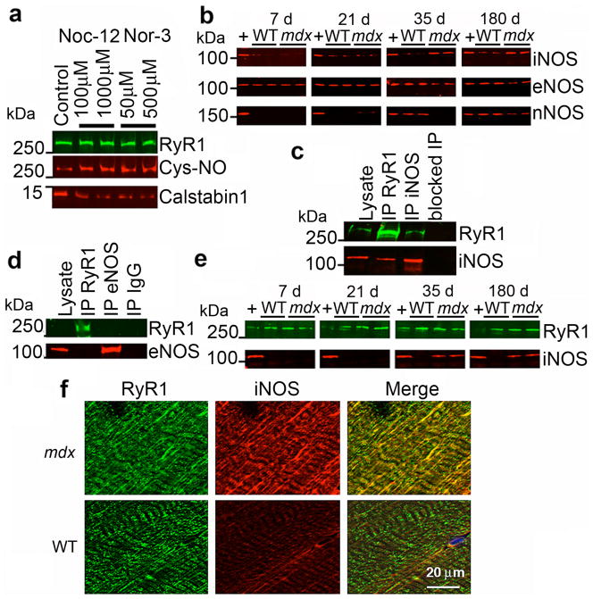
Figure 2. iNOS co-immunoprecipitates and co-localizes with RyR1 and S-nitrosylation of RyR1 depletes the channel of calstabin1.
(a) In vitro S-nitrosylation of skeletal SR microsomes with NO donors Nor-3 or Noc-12 results in depletion of calstabin1 from immunoprecipitated RyR1. (b) Immunoblot of expression of three NOS isoforms (iNOS, eNOS, and nNOS) from WT and mdx whole muscle lysates at the indicated ages. (c) Co-immunoprecipitation of RyR1 and iNOS. 50 μg of mdx EDL lysate was used as positive control. RyR1 and iNOS separately immunoprecipitated from 250 μg of mdx muscle lysate and probed for RyR1 and iNOS. Antibody against RyR was pre-incubated with 100-fold excess antigenic peptide prior to immunoprecipitation (blocked IP RyR). (d) Immunoprecipitation-immunoblotting of RyR1 and eNOS from mdx lysate. IgG control immunoprecipitation shown at right. (e) RyR1 was immunoprecipitated from WT and mdx EDL lysates at indicated ages and immunoblotted for RyR1 and iNOS. (f) Immunohistochemistry showing co-localization of RyR1 and iNOS in murine skeletal muscle (EDL) from mdx but not WT mice. Scale bar in lower right panel applies to all six panels.

