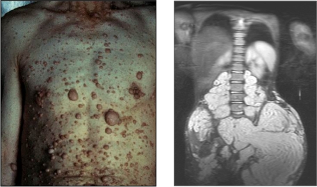
Figure 1.
Examples of cutaneous and plexiform neurofibromas. Cutaneous neurofibromas growing on the chest and abdomen and an magnetic resonance image of a large plexiform neurofibroma compressing the spinal column. Photographs courtesy of the Children's Tumor Foundation; www.ctf.org.

