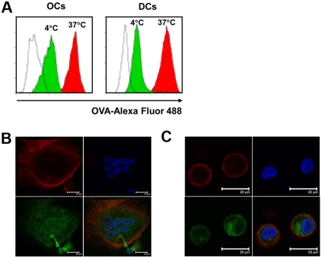
Figure 6.
Uptake of soluble antigens by OCs. OCs and immature DCs were incubated with OVA–Alexa Fluor 488 at a concentration of 1 mg/mL at 37°C or 4°C for 30 minutes. After washing and fixation, cells were analyzed by flow cytometry and confocal microscope. (A) Flow cytometric analysis of the uptake of OVA–Alexa Fluor 488 by OCs and DCs. Unfilled curves show cells incubated without OVA–Alexa Fluor 488, green curves show cells incubated with OVA–Alexa Fluor 488 at 4°C, and red curves show cells incubated with OVA–Alexa Fluor 488 at 37°C. Images of (B) OCs or (C) DCs by confocal microscopy. Red shows staining with anti-CD11c antibody, green shows staining with OVA–Alexa Fluor 488, and blue shows nuclei stained with DAPI. Scale bar represents 20 μm. Representative results from 1 of 3 experiments performed are shown.

