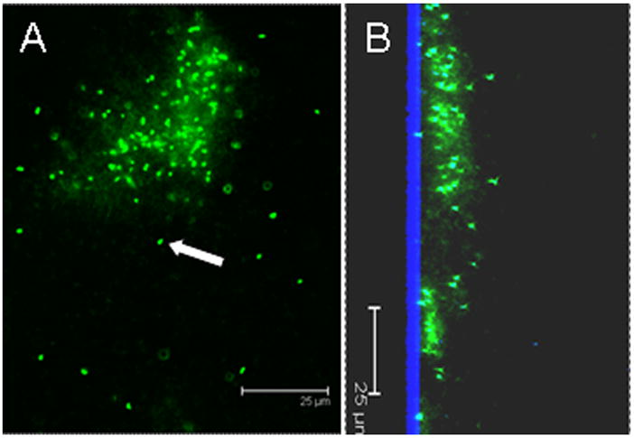
Figure 2.

Confocal micrograph of a P. aeruginosa PAO1 biofilm grown on a glass substratum demonstrating extracellular DNA within the biofilm matrix surrounding the bacteria. A) plan view, and B) saggital section with the glass substratum is shown in blue by reflected light. The biofilm was stained with the DNA stain Syto9. The cells (an example of a non-matrix enclosed cell is shown with white arrow) were slightly overexposed to show the more diffuse signal from the extracellular DNA (eDNA) between the individual bacteria. This image demonstrates: 1) the close proximity of the cells within the biofilm; 2) that within biofilms, which are relatively quiescent (unlike laboratory planktonic cultures which are continually mixed), the cells have more time to interact (i.e. for conjugation), and 3) there is a pool of eDNA surrounding the cells which provides structural stability as well as serving as a source for transformation. Scale bars = 25 μm.
