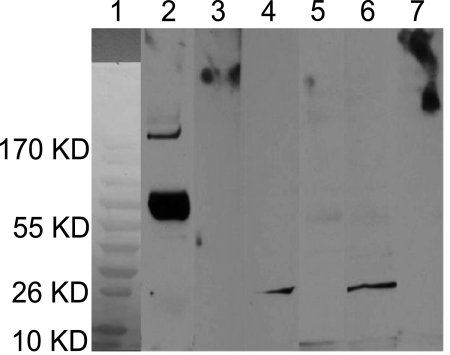
Figure 4.
Immunoblot using conjunctival proteins recovered from cases and controls. Total pooled proteins (10 μg) were transferred to nitrocellulose membranes and probed with an anti-MMP7 primary (1°) antibody. Bound antibody was detected with a secondary (2°) antibody conjugated to HRP (anti-mouse IgG-HRP) and detected by ECL. Control lanes were probed with 2° antibody alone. Molecular weight markers were used in lane 1 to estimate the size of unknown bands from pooled samples. Lanes 2 and 3 were loaded with 0.1 ng of recombinant human MMP7 (rhMMP7) as a positive control (∼70 kDa when probed with anti-MMP7). Probing with 2° antibody alone (lane 3) elicited no reactivity. A band at ∼26 kDa was visible in lanes 4 (pooled cases) and 6 (pooled controls) when probed with monoclonal anti-MMP7 antibody. No bands were visible in lanes 5 (pooled cases) and 7 (pooled controls) when probed with 2° antibody alone.

