Figure 2.
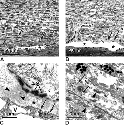
(A, B) Semithin sections of the TM and outflow loops (Schlemm's canal equivalent in the cow; asterisks) of a 6-month-old calf eye (A) and an untreated control Braford cow eye (B). (A) In the calf eye, the inner TM is more reticular than lamellar. The outer trabecular cells are connected to the outflow loops by smaller and more elongated TM cells (arrows) than those of the remaining reticular meshwork. There is no plaque material under the inner wall of the outflow loop (stage 0). (B) In the untreated Braford cow eyes, the TM also is reticular. There are only few elongated cells in the subendothelial region of the loops. Directly under the endothelium are some plaques (stage 1, arrows), but they are sparsely located. (C, D) Electron micrographs of untreated Braford cow control eyes. (C) In the subendothelial region (asterisk), fine fibrils (arrowheads) span the area from the elastic fiber sheath (S) to the endothelium. Accumulations of these connecting fibrils appear as loosely arranged plaques. At places of fibril attachment, the endothelium forms dense bands (arrows). V, giant vacuole. (D) TM cells between the collagen and elastic fibers show nearly no basement membranes. Within their cytoplasm are numerous glycogen granules (arrows). Elastic fibers contain electron light material within the osmiophilic core and a sheath of fine fibrillar material (arrowheads).

