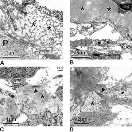
Figure 4.
Electron micrographs of the subendothelial region of steroid-treated eyes. (A) Irregularly arranged basement membrane-like material (asterisks) can be seen adhering to the fine fibrillar subendothelial plaques (p). (B) Fibrillar material within the plaques (asterisks) is more densely packed than in the controls, and fibrils are embedded in a more osmiophilic ground substance. (C) Fine fibrils (asterisk) of the plaques often adhere to the sheath of the subendothelial elastic fibers (arrowhead). (D) Fibrils of the plaques are cross-linked (arrowhead). Occasionally, they are in contact with basement membrane-like material (asterisk).

