Figure 5.
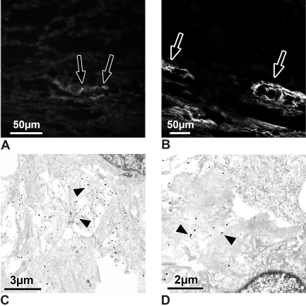
(A, B) Light micrographs of sections through outflow loops, immunohistochemically stained for type VI collagen. (A) Calf eye. There are only small dots (arrows) of type VI collagen staining under the endothelium of the outflow loop's inner wall. (B) Steroid-treated eye. Extracellular material surrounding the loops stains intensely for type VI collagen (arrows) in treated and in contralateral control eyes. (C, D) Electron micrographs of the inner wall of outflow loops immunogold-labeled for type VI collagen. (C) Contralateral control eye. Note that the fine fibrils in the subendothelial region are labeled intensely (arrowheads) for type VI collagen. (D) Steroid-treated eye. Where the plaques contained cross-linked fibrils, only those fibrils that entered the plaques are labeled (arrowheads).

