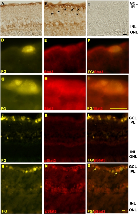
Figure 1.
Stat3 protein increased in the glaucomatous retina and in glaucomatous RGCs, and phospho-Stat3 was also increased in glaucomatous RGCs. (A) In the normal retina, positive staining for Stat3 was seen in the inner nuclear layer (INL) and GCL. (B) The expression of Stat3 was higher in the GCL in the high-IOP retina. (C) Negative control. (E) Low levels of Stat3 immunoreactivity were seen in Fluorogold-labeled RGCs in a retina with normal IOP. (G–I) In retinas with high IOP, increased Stat3 expression was seen in Fluorogold-labeled RGCs. (K–N) Phosphorylated-Stat3 was clearly increased in the GCL, including in RGCs (O compared with L). FG, Fluorogold. Arrow: retinal ganglion cell; Arrowhead: cell processes. Scale bars: 20 μm.

