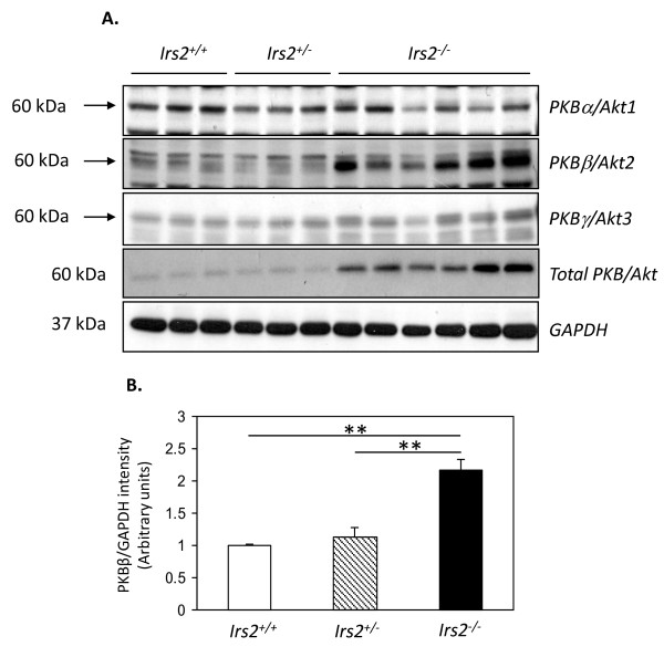
Figure 5.
Specific increase in PKBβ/Akt2 is detected in Irs2-/- kidney. (A) Twenty μg of protein lysates from Irs2+/+ (n = 3), Irs2+/- (n = 3) and Irs2-/- (n = 6) mice were separated on 10% SDS-PAGE and probed by Western blotting with anti-PKBα/Akt1, PKBβ/Akt2, PKBγ/Akt3 and anti-PKB/Akt antibodies. GAPDH was used as a loading control. (B) Band intensities were quantified using Scion Image software. The intensity ratio of anti-PKBβ/GAPDH is shown. ** p < 0.01.

