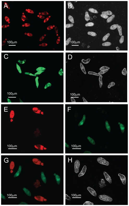
Figure 1. Fluorescein diacetate (FDA) and propidium iodide (PI) can be used to differentially quantify schistosomula viability.
Mechanically-transformed schistosomula were prepared, heat-killed (dead) or left untreated (live) and stained with FDA, PI or both fluorophores according to the Methods . Epi-fluorescent and plane polarized microscopy was used to visualize uptake of fluorophores and examine schistosomula morphology. (A) Dead schistosomula stained with PI and detected by a rhodamine filter (536 nm), (B) Dead schistosomula visualized by polarized light, (C) Live schistosomula stained with FDA and detected by a FITC filter (494 nm), (D) Live schistosomula visualized by polarized light (E) Mixtures of dead and live schistosomula simultaneously stained with PI/FDA and detected by a rhodamine filter, (F) Mixtures of dead and live schistosomula simultaneously stained with PI/FDA and detected by a FITC filter, (G) Differential detection of PI-positive dead and FDA-positive live schistosomula by superimposition of both 536 nm and 494 nm epifluorescent spectra, (H) Differential morphology of dead and live schistosomula detected by polarized light.

