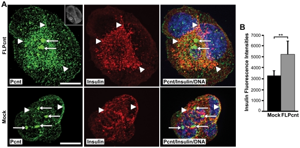
Figure 2. Overexpression of pericentrin in insulinoma cells caused an increase in the granular intracellular insulin staining.
A. MIN6 cells were transfected with a FLAG-tagged pericentrin expression construct for 48 hr and immunostained with pericentrin (green) and insulin (red) antibodies (FLAG upper right in pericentrin box); arrows indicate the centrosome and arrowheads point to the granular cytoplasmic staining. Yellow shows overlay of pericentrin and insulin immunostaining; nuclei are blue; scale bar represents 10 µm. B. Quantitation of insulin fluorescence intensities (** p<0.01). All error bars represent ± SEM. All data shown are representative of multiple independent experiments.

