
Fig. 2.
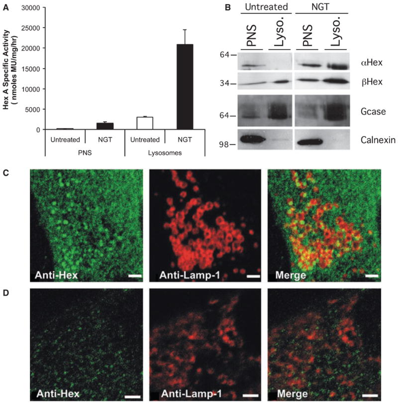
Inhibitors of Hex, NGT and PYR increase levels of mutant Hex A in late-onset Tay-Sachs disease and Sandhoff disease patient fibroblasts. (A) Comparison of MUGS activity (nmol·mg of total cell protein−1·h−1) in the postnuclear supernatant (PNS) and lysosome-enriched (Lysosomes) fractions from untreated (open bar) and NGT- (0.9 mM) treated (filled bar) ATSD (αG269S) fibroblasts. (B) Western blots comparing the levels of mature αG269S- (α Hex) and wild-type β-subunits (β Hex) of Hex; a lysosomal marker, glucocerebrosidase (Gcase), and an ER marker, calnexin, in the PNS and lysosomal fractions (Lyso.) from untreated and NGT-treated ATSD cells. (C, D) Increased colocalization (merge-yellow) of mutant βR505Q-containing Hex isozymes in an ASD patient cell line, visualized with anti-β-subunit Hex (anti-Hex) IgG (stained green), or with lysosome associated membrane protein-1 (Lamp-1) visualized with anti-Lamp-1 (anti-Lamp) IgG (stained red). (C) Cells were treated with PYR (3.0 μg·mL−1) or (D) with the solvent (ethanol) used to dissolve PYR. Scale bars (2 μM) are provided in the bottom right-hand corner of panels.