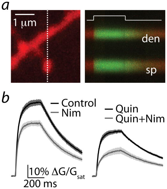
Figure 6. Voltage-steps reveal L-type Ca channels in MSN spines.
(a) left, 2PLSM image of a striatopallidal MSN dendrite filled with Alexa Fluor 594 and Fluo 5F. right, Red and green fluorescence in the spine head (sp) and neighboring dendrite (den) measured in line scan over the region indicated by the dashed line at left. The increase in Ca-dependent green fluorescence was evoked by a 300 ms voltage step from −70 mV to 0 mV at the time indicated by the white line.
(b) left, mean ± SEM (shaded traces) are shown for responses evoked under control conditions (n=22, black) and in the presence of nimodipine (n=21, gray).
right, mean ± SEM for responses evoked in the presence of quinpirole (n=20, black) or quinpirole + nimodipine (n=20, gray).

