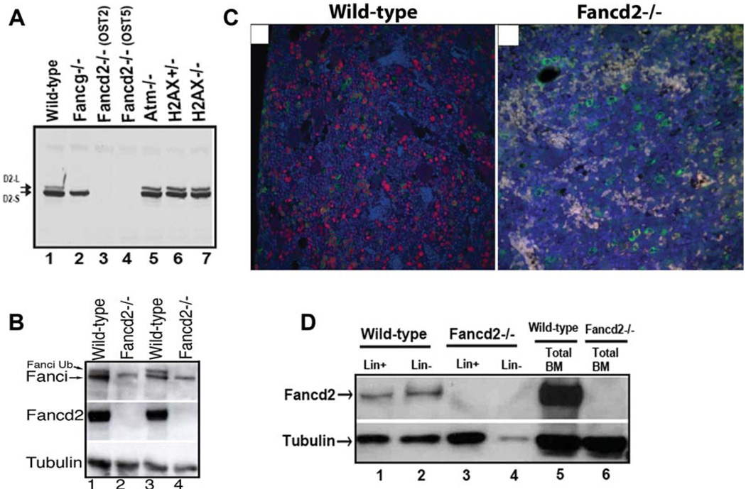
Figure 1.
Fancd2 expression in murine tissues. (A): Western blot of Fancd2 in splenocytes with indicated genotypes. Note that Fancd2 protein expression is deficient in both OST2 and OST5 mouse lines (lanes 3, 4). (B): Western blot of Fanci and Fancd2 in bone marrow cells from four different mice with indicated genotypes. (C): Detection of Fancd2 in the bone marrow in the bone sections using the HistoRX fluorescence imaging system. Representative examples of fluorescent staining for Fancd2 (red) and myeloperoxidase (green) on wild-type and Fancd2−/− paraffin embedded bone sections are shown. DAPI stain (blue) highlights the total nuclei. Note that Fancd2 staining (red) is observed in subset of the myeloperoxidase stained myeloid cells (green) as well as in the myeloperoxidase negative cells in the wild-type bone marrow. Magnification ×400. (D): Western blot of Fancd2 in fractionated Lin+ and Lin− BM cells from wild-type and Fancd2−/− mice. Abbreviations: BM, bone marrow; Lin−, lineage negative; Lin+, lineage positive.

