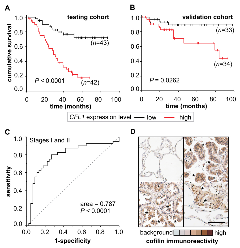
FIGURE 2. Biomarker performance in early stage NSCLC patients.
(A) Kaplan Meier plot are shown for patients in stages I and II (n=85) in the original cohort (testing cohort) stratified by CFL1 expression level and (B) in an independent cohort (validation cohort) obtained from a different set of published NSCLC microarray data (n=67). (C) Biomarker performance estimated by Receiver Operating Characteristic (ROC) analysis. (D) Representative immunohistochemical (IHC) analysis of cofilin immunocontent in tumor biopsies. Healthy human alveolar tissue obtained from tumor margins is mostly negative to cofilin IHC staining (upper left). High staining for cofilin is found within the neoplasic lung cells (asterisks). Original magnification ×200; scale bar = 100 µM.

