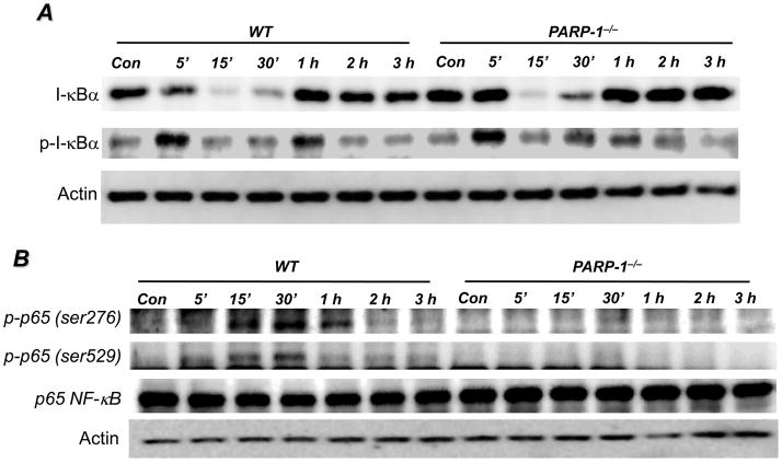
Fig. 2. Effects of PARP-1 gene deletion on phosphorylation and degradation of I-κBα and phosphorylation of p65 NF-κB in SMCs upon LPS treatment.
WT or PARP-1−/− SMCs were treated with 1 μg/ml LPS for different time intervals or left untreated (Con). (A) Proteins extracts were subjected to immunoblot analysis with antibodies to I-κBα or phosphor-I-κBα at Ser32/Ser36. (B) The same extracts were subjected to immunoblot analysis with antibodies to phospho-p65 NF-κB at Ser276 or Ser529, total p65 NF-κB, or actin.

