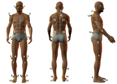
Figure 1.
This figure shows the anatomic points that were visualized. Footnote: tragus (1); medium point, acromion (2); anterior-superior iliac spine (ASIS) (3); femur, greater trochanter (4); knee, articular line (5); patella, medium point (6); tibia tuberosity (7); lateral malleoli (8); medial malleoli (9); medium point between second and third metatarsus (10); spinal process of C7 (11) and T3 (12); scapula, inferior angle (13); posterior-superior iliac spine (14); leg, point a medial line (15); calcaneum tendon between malleolus (16); and calcaneum (17).

