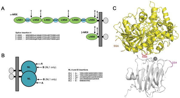
Figure 1. NRX and NL alternative splicing.
(A) NRXs are transcribed from two independent promoters for each gene resulting in the longer α-NRXs and shorter β-NRXs. α-NRXs contain three repeats, each of which consists of an EGF domain flanked by two LNS domains. α-NRX and β-NRXs share the LNS 6 domain, a transmembrane region and a short cytoplasmic domain. The arrows indicate the alternative splicing sites in each molecule. The amino acids sequences for the insert at splice site 4 of each β-NRX are also shown. (B) NLs 1, 2, and 3 contain an extracellular domain, which forms homodimers, a stalk region, transmembrane and cytoplasmic domains. All three NLs undergo splicing at site A and NL1 only can also be spliced at site B, resulting in 10 different NL variants. The amino acid sequences for the inserts introduced as a result of splicing are shown. (C) Structure of a β-NRX1Δ4/NL1Δ complex with β-NRX1Δ4 shown in silver and NL1Δ in yellow (PDB ID 3B3Q) (Chen et al., 2008). The SS4 insertion point for β-NRXs is shaded in magenta, the SSA insertion point for NLs is shaded in orange and the SSB insertion point for NL1 in red.

