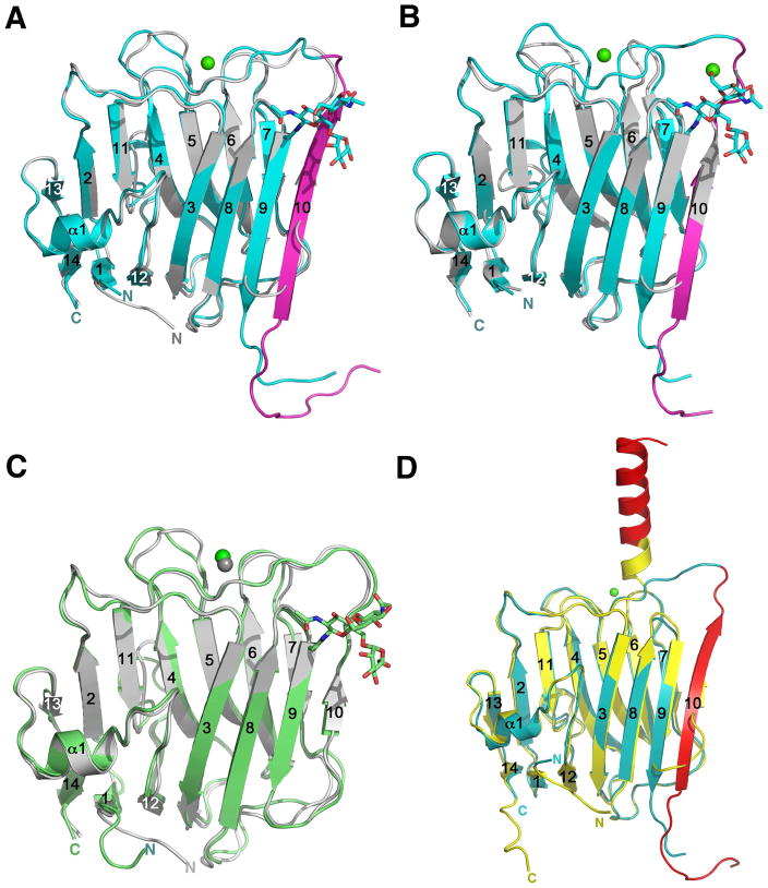
Figure 3.
Structural rearrangements take place to accommodate the splice insert 4. (A) Superposition of the β-NRX1Δ4 structure (PDB ID 3BOD) in silver and the β-NRX1+4 structure in cyan. The SS4 insert is highlighted in magenta. (B) Superposition of the β-NRX2Δ4 structure (PDB ID 3BOP) in silver and the β-NRX2+4 structure in cyan. The SS4 insert is highlighted in magenta. (C) Superposition of the β-NRX1Δ4 structure (PDB ID 3BOD) in silver and the glycosylated β-NRX3Δ4 structure in green. (D) Superposition of the new β-NRX1+4 structure in cyan with the non-glycosylated β-NRX1+4 structure (PDB ID 2R1B) (Shen et al., 2008) in yellow. The SS4 is shown in red in both structures, highlighting identical sequences adopting completely different secondary structures.

