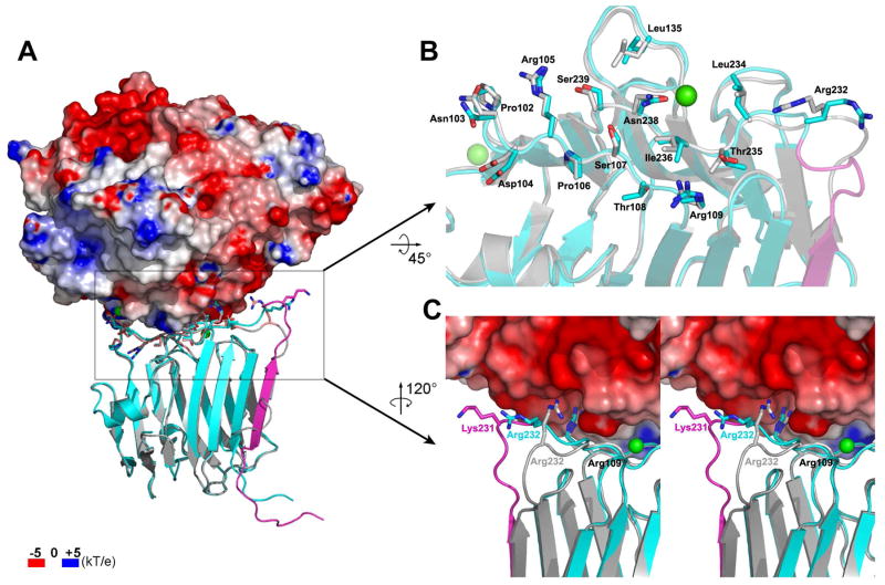
Figure 4.
Structural effects of the SS4 insertion on the NL binding site. (A) The β-NRX1+4 structure (cyan) is superposed onto the β-NRX1Δ4 (silver) from the β-NRX1Δ4/NL1Δ complex structure (PDB ID 3BIW). The SS4 insert is shown in magenta in the β-NRX1+4 structure. The electrostatic potential surface is shown for the NL1 molecule in the complex with the indicated color scale. (B) Close-up view of the NL binding interface of β-NRX1Δ4 with the β-NRX1+4 structure superposed. The β-NRX1Δ4 interface residues with a buried surface area greater than 5Å2 in the complex structure with NL1 and the same residues in β-NRX1+4 are represented. The colorcoding is the same as in (A). (C) Close-up view of the part of the β10–β11 loop that is the most variable upon SS4 insertion.

