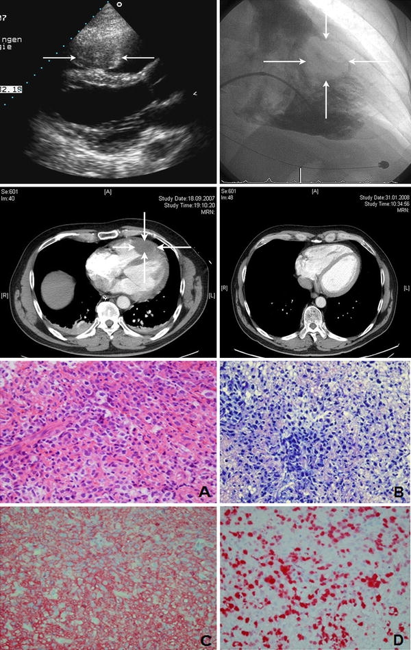
Fig. 1.

Upper left panel: transthoracic echocardiogram shows an echodense structure in the right ventricle (arrows). Upper rightpanel: right ventricular angiography demonstrating an intraventricular filling defect after injection of contrast medium. Middle panel: contrast-enhanced computed tomography demonstrating an intraventricular filling defect after the injection of contrast material at the time of diagnosis (left) and after completing the last course of chemotherapy (right). Lower panel: histologic examination of the cardiac tumor biopsy showed sheets of large lymphoid cell admixed with small reactive lymphocytes [hematoxylin and eosin (a) and Giemsa (b) stains; ×400 magnification]. Immunohistochemistry demonstrated CD20 expression (c) on neoplastic cells. The proliferation rate was up to 80% (d ×400 magnification)
