
Fig. 2.
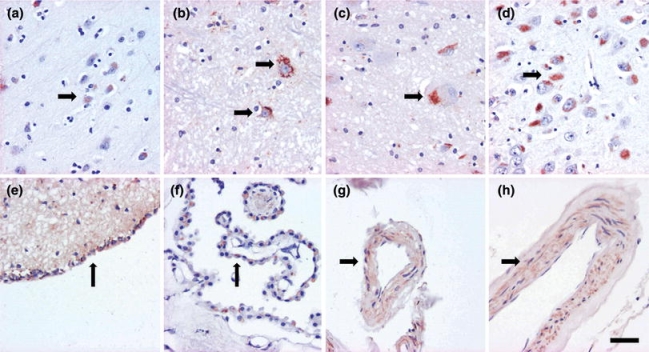
Note the brown cytoplasmic amyloid-β labeling seen when applying the 4G8 antibody in neurons and glial cells (a, b, c, d), ependyma (e), choroid plexus (f) and vessel walls (g). This labeling was not utilized in the classification. Magnification ×400, scale bar 10 μm