
FIGURE 2.
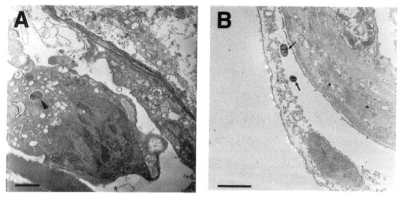
Analysis of C. pneumoniae-infected cells in AD blood vessels. Panel A: Blood vessel from the hippocampus from an AD brain showing an RB (arrowhead) within the cytoplasm of a presumptive monocyte/macrophage in the vessel lumen. Panel B: Blood vessel from the temporal cortex from an AD brain demonstrating two C. pneumoniae organisms (arrows) within the disrupted cytoplasm of a detached pericyte or perivascular macrophage on the abluminal surface of the vessel. Bars= 1 μm