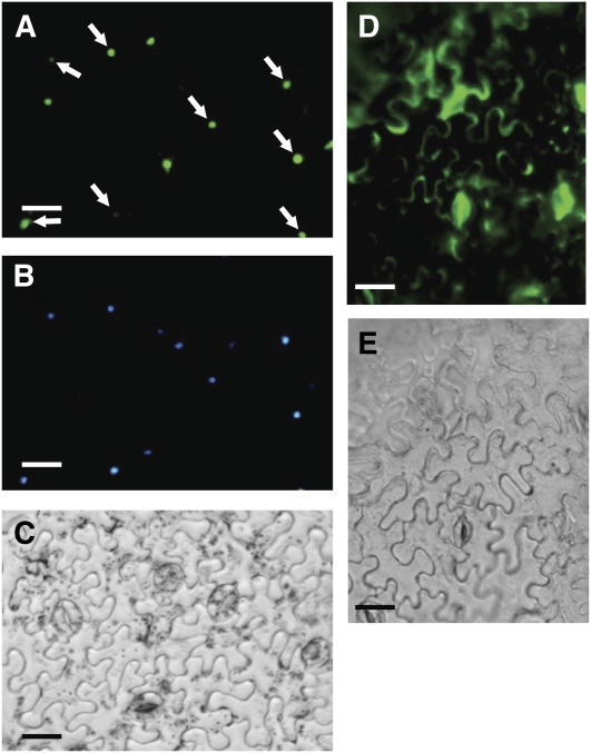
Figure 6.
Nuclear Localization of EOBII.
(A) Localization of the EOBII:GFP fusion protein in the leaf epidermis.
(B) Nucleus stained with 4',6-diamidino-2-phenylindole.
(C) Bright field.
(D) and (E) Fluorescence of nonfused GFP used as a control and bright field of leaf epidermis, respectively. Arrows indicate nuclei with 4',6-diamidino-2-phenylindole and GFP cofluorescence.
Bars = 25 μm.
[See online article for color version of this figure.]

