Figure 6.
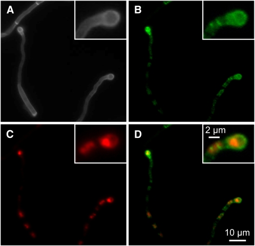
Localization of Sho1-GFP and Msb2-mCherry in Appressoria.
(A)to (D) SG200sho1GFP/otef:msb2mCherryHA was sprayed with 100 μM 16-hydroxyhexadecanoic acid on paraffin wax and incubated as described in Figure 5B. A section displaying two appressoria is analyzed. The appressorium on the right-hand side is magnified in the inset.
(A) Calcofluor staining.
(B) Visualization of Sho1-GFP (green).
(C) Visualization of Msb2-mCherry (red).
(D) Overlay of (B) and (C).

