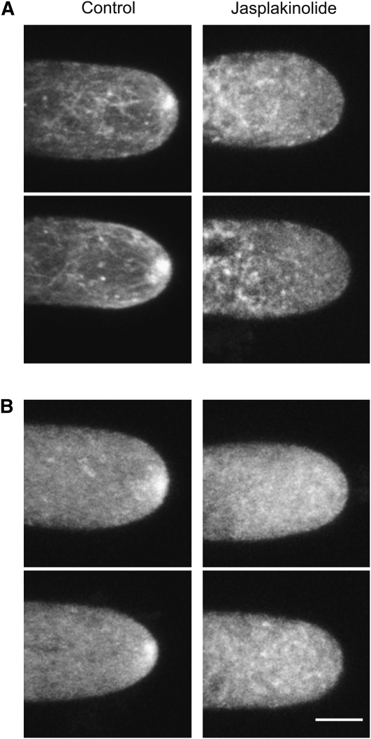
Figure 8.
Stabilization of the Actin Cytoskeleton Delocalizes Myosin XI from the Apex of the Cell.
(A) Two representative images of F-actin distribution visualized with Lifeact-mEGFP in cells treated with DMSO (left panels) or with 20 μM Jasplakinolide (right panels) for 30 min.
(B) Two representative images of 3xmEGFP-myoXIa cells treated with DMSO (left panels) or with 20 μM Jasplakinolide (right panels) for 30 min. Images are the maximal projection of five focal planes acquired at 1-μm intervals from the medial section of the cells. Bar = 5 μm.

