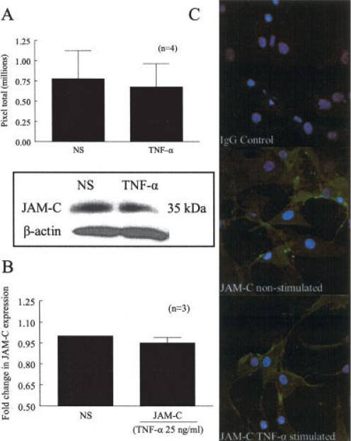
Figure 2.
JAM-C expression in RA ST fibroblasts. A, Mean and SEM total number of pixels showing JAM-C expression in RA ST fibroblasts left untreated or cultured with tumor necrosis factor α (TNFα) for 24 hours. Western blots showed a 35-kd protein band consistent with the molecular weight of JAM-C; however, no differences were observed after stimulation with TNFα. B, Mean and SEM fold change in expression of JAM-C on the cell surface of RA ST fibroblasts left untreated or cultured with TNFα, determined by cell surface enzyme-linked immunosorbent assay. Surface expression of JAM-C on RA ST fibroblasts did not change after stimulation with TNFα. C, Surface expression and localization of IgG control and JAM-C on RA ST fibroblasts, examined using immunofluorescence and confocal microscopy. JAM-C was expressed throughout the surface, and no difference in expression was noted between adjacent fibroblasts in the presence or absence of TNFα. NS = nonstimulated (see Figure 1 for other definitions).

