
FIGURE 4. Pericontusional AQP4 expression following TBI.
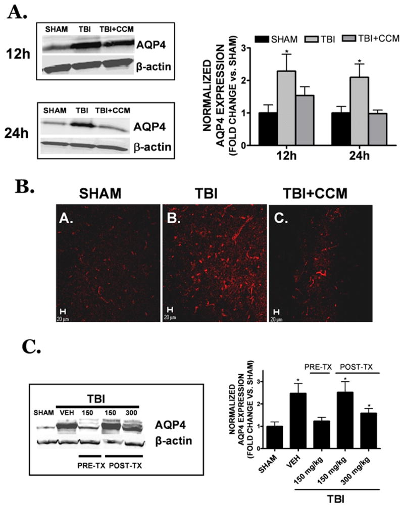
(A) Curcumin (150 mg/kg) was administered to mice at 15 minutes prior to injury and AQP4 expression was assessed 12h or 24h later by Western blotting. AQP4 expression was normalized to β-actin to control for equal protein loading. (B) Representative low power (20x objective) images of AQP4 immunoreactivity within the peri-contusional cortex following (A) sham, (B) TBI or (C) TBI + curcumin pre-treatment (150 mg/kg administered 0.5h prior to TBI). Data are representative of n=4 animals/group. Scale bar = 20 μm. (C) Representative Western blot of AQP4 expression (left) at 24h post-TBI following vehicle, 150 mg/kg curcumin pre-treatment (PRE-TX), or 1h post-treatment (150 or 300 mg/kg, POST-TX). For both panels, quantification of Western blotting (5-7 mice/group in each treatment condition) was performed by densitometry analysis. Data were represented as the mean ± SEM at each time point and expressed as fold change vs. sham. Data were analyzed using One-Way ANOVA followed by Student Newman Keul’s post-hoc test (*p<0.05 vs. sham).