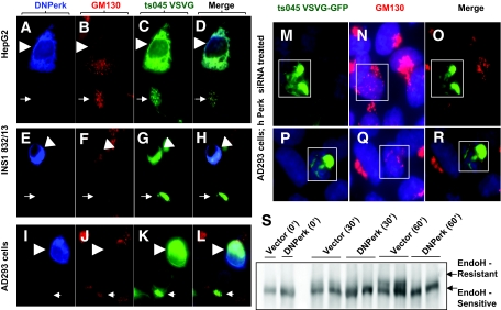
FIG. 2.
An acute ablation of Perk function leads to an ER to Golgi anterograde trafficking defect. IHC in HepG2 cells (A–D), INS1 (832/13) (E–H), AD293 cells (I–R), and EndoH resistance assay in AD293 cells (S). A–L: Cells were cultured on Lab-Tec (Nalge Nunc) chamber slides prior to transfection with DNPerk and ts045 VSVG-GFP followed by IHC analysis of detergent-permeabilized cells (0.2% Triton X 100 in 1 × PBS for 10 min at room temperature). Arrowheads show a DNPerk and ts045-VSVG-GFP cotransfected cell following a 16-h incubation at the restrictive temperature and a 15-min shift to the permissive temperature. Arrows indicate an adjacent control cell transfected with only ts045-VSVG-GFP. A–D: HepG2 cells were cotransfected with c-myc tagged DNPerk (blue, A) and ts045-VSVG-GFP (green, C) and stained for the Golgi marker GM130 (red, B). The merged image is seen in D. E–H: INS1 (832/13) cells were cotransfected with c-myc–tagged DNPerk (blue, E) and ts045-VSVG-GFP (green, G) and stained for the Golgi marker GM130 (red, F). The merged image is seen in H. I–L: AD293 cells were cotransfected with c-myc–tagged DNPerk (blue, I) and ts045-VSVG-GFP (green, K) and stained for the Golgi marker GM130 (red, J). The merged image is seen in L. M–R: IHC in AD293 cells. Boxed areas in M and P show cells cotransfected with ts045 VSVG-GFP (green) and Perk siRNA after an 8-h incubation at the restrictive temperature followed by a 15-min shift to the permissive temperature. Human Perk mRNA was reduced by 41% at 8 h posttransfection of the siRNA. Staining for the Golgi marker GM130 shows a dispersed morphology in red (N and Q) and the merged images are seen in O and R. S: ts045-VSVG-GFP was cotransfected with either empty vector (Vector) or DNPerk into AD293 cells. Cells were incubated overnight at the restrictive temperature to allow the VSVG to accumulate in the ER (0′). Cells were then shifted to the permissive temperature to allow the protein to traffic to the Golgi. Cells were harvested for EndoH treatment and Western blots at the indicated time points. The higher MW form (upper arrow) is the EndoH-resistant Golgi form, while the lower arrow indicates the ER-retained EndoH-sensitive form. (A high-quality digital representation of this figure is available in the online issue.)

