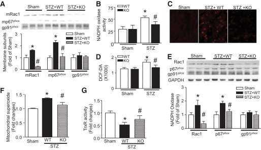
FIG. 1.
Effects of Rac1 knockout on NADPH oxidase and ROS production. Rac1-ko mice (KO) and their WT littermates were injected with STZ. Two months later, NADPH oxidase activation and expression and ROS production in heart tissues were measured. A: Translocalization of Rac1 and p67phox to the membrane. The protein levels of Rac1 (mRac1) and p67phox (mp67phox) were decreased in the membrane fractions of Rac1 KO compared with WT diabetic hearts. The top panel is the representative Western blot for membrane mRac1, mp67phox, and gp91phox from three out of five to six different hearts in each group, and the lower panel is the quantification of mRac1, mp67phox, and gp91phox. NADPH oxidase activity (B), superoxide production (C), and H2O2 production (D) were decreased in diabetic Rac1 KO compared with WT hearts. C is the representative DHE staining (Red signal) for superoxide production from five to six different hearts in each group. E: Rac1, p67phox, and gp91pho protein expression. The protein levels of Rac1 and p67pho were decreased in Rac1 KO compared with WT diabetic hearts. The top panel is the representative Western blot for Rac1, p67phox, and gp91phox from three out of five to six different hearts in each group and the lower panel is the quantification of Rac1, p67phox, and gp91phox. F: Mitochondrial superoxide production was increased in WT diabetic hearts, which was significantly decreased in Rac1 KO hearts. G: Thioredoxin reductase activity was preserved in Rac1 knockout diabetic hearts. Magnification ×40. Data are means ± SD, n = 5–8. *P < 0.05 vs. sham; #P < 0.05 vs. STZ in WT. (A high-quality digital representation of this figure is available in the online issue.)

