FIG. 4.
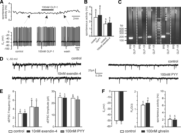
PPG neurons are not sensitive to gut peptides GLP-1 and PYY. A: Perforated patch clamp recording in current clamp demonstrating the lack of effect of 100 nmol/l GLP-1 on PPG cell activity. The top trace shows the instantaneous firing frequency over the course of the recording, and the bottom traces show brief periods of the original recording at time points indicated by arrows. B: Mean firing frequency in the absence and presence of 100 nmol/l GLP-1 or 100 nmol/l of the GLP-1 receptor agonist exendin-4. C: Typical single-cell RT-PCR analysis for PPG and the GLP-1 receptor (GLP-1R) for three PPG neurons and controls. The 2% agarose gel demonstrating that the 186-bp PCR product for PPG and the 292-bp PCR product for GLP-1R can be obtained from brainstem cDNA (BS 1:1,000 dilution; positive control) with the primers specified in Table 1. In contrast, cytoplasm extracted from single cells showing Venus fluorescence (1–3) was only positive for PPG, but not GLP-1R. PIP, pipette solution without cytoplasm extracted from cell (negative control). D: Neither exendin-4 nor the gut peptide PYY had any effect on spontaneous EPSCs in PPG neurons. Left: Typical voltage-clamp recordings in the absence or presence of exendin-4. Right: Typical recordings in the absence or presence of PYY. E: Mean data from recordings as shown in B for frequency and amplitude of spontaneous EPSCs. F: The peptide ghrelin (100 nmol/l), released from stomach as orexinergic signal, had no effect on membrane potential (EM), input resistance (RM), or action potential frequency. Numbers of cells tested (n) are given above the bars.

