



Abstract
Leptospirosis, a zoonosis occurring worldwide, has a broad spectrum of clinical manifestations. Recently, various countries observed an increase of severe anicteric cases. In Austria and Germany, growing numbers of imported cases are notified in addition to autochthonous infections. The aim of this study was to assess whether imported and autochthonous cases differ in clinical manifestations and outcome. We retrospectively analyzed 24 imported and 35 autochthonous cases treated in six infectious disease units between 1998 and 2008. To compare disease severity, patients were classified according to established independent risk factors for fatal outcome. Although severe leptospirosis (i.e., presence of ≥ 1 independent risk factors for death) occurred in similar proportions of imported (67%) and autochthonous (86%) infections (P = 0.1), imported cases were significantly fewer icteric (13% versus 69%; P < 0.0001). In conclusion, an increasing incidence of severe anicteric imported cases of leptospirosis should be anticipated with rising global travel activities.
Introduction
Leptospirosis, probably the most common zoonosis worldwide,1 has gained increasing attention as an emerging infectious disease of global importance in recent years.2,3 Its clinical manifestation is divers,2,4 ranging from asymptomatic infections to severe and potentially fatal disease complicated by septic shock and organ failure. This broad clinical spectrum is presumably one of the main reasons why management of this spirochaetosis remains a challenge for clinicians. Because of the wide variety of mostly unspecific clinical and laboratory findings, a multitude of differential diagnoses comes into account. Among these are dengue fever, acute human immunodeficiency virus (HIV), and hantavirus infections, rickettsial diseases, brucellosis, malaria, and other infections of significant public health importance such as yellow fever and various viral hemorrhagic fevers.2,3,5
In most cases, leptospirosis is a subclinical or undifferentiated self-limited febrile illness.6,7 In about 5–10% of cases, however, the disease takes a severe course with the potential of multi-organ dysfunction syndrome (MODS) with a high risk of fatal outcome. Jaundice has traditionally been considered as an indicator of complicated forms, the most severe being Weil's disease with the triad of jaundice, acute renal failure (ARF), and hemorrhages and a mortality rate of 5% to 15%.2,8 In recent years, however, an increasing number of severe anicteric forms has been observed in various countries like China, Korea, Nicaragua, Peru, and India.9–14 In these cases, serious pulmonary manifestations with severe pulmonary hemorrhage syndrome (SPHS) and acute respiratory distress syndrome (ARDS) predominated. The mortality rate of up to 50% of these manifestations appeared to be much higher than in Weil's disease. Hence, jaundice alone can no longer be considered an accurate marker of disease severity.15 There is, however, currently no consistent definition of what characterizes “severe forms” of leptospirosis, although several factors associated with an increased risk of fatal outcome (therefore being prognostically relevant) have been identified over the past decade (Table 1).8,16–26
Table 1.
Independent risk factors for death, severity score of leptospirosis (used for risk factor analysis as shown in Table 2) and corresponding criteria of (severe) sepsis and multi-organ dysfunction syndrome (MODS)*
| Independent risk factor for fatal leptospirosis | Reference | Odds ratio | Score | Corresponding criteria of (severe) sepsis and MODS |
|---|---|---|---|---|
| Age > 40 years | 17 | 4.38 | 1 | |
| 23 | 2.2 | |||
| WBC count on admission > 12.9/nL | 18 | 2.5 | 1 | WBC count > 12/nL |
| Altered mental status | 17 | 9.12 | 1 | Altered mental status |
| 26 | 8.9 | |||
| 24 | – | |||
| Hypotension/shock (need of vasoactive substances to maintain arterial blood pressure ≥ 80 mm of Hg after adequate fluid replacement) on admission | 21 | 6.0 | 1 | Systolic blood pressure ≤ 90 mm of Hg or mean arterial pressure ≤ 70 mm of Hg for at least 1 hour despite adequate volume resuscitation or the use of vasopressors to achieve the same goals |
| 19 | 10.3 | |||
| 20 | – | |||
| Repolarization abnormalities on ECG | 18 | 5.9 | 1 | |
| Arrhythmias | 17 | 2.83 | 1 | |
| Oliguria | 18 | 9.0 | 1 | Urine output < 0.5 mL/kg of body weight/hour or ARF |
| 17 | 5.28 | |||
| 16 | 8.98 | |||
| 19 | 8.8 | |||
| 25 | – | |||
| 20 | – | |||
| 23 | 3.0 | |||
| BUN > 324 mg/dL (= 54 mmol/L) | 17 | 3.86 | 1 | See oliguria |
| Serum creatinine > 4.0 mg/dL (> 354 µmol/L) | 17 | 2.82 | 1 | See oliguria |
| 21 | 10.6 | |||
| 23 | 2.3 | |||
| Hyperkalemia > 5.0 mmol/L on admission | 17 | 8.27 | 1 | |
| 22 | – | |||
| 21 | 19.9 | |||
| 26 | 4.2 | |||
| 19 | 5.9 | |||
| Dyspnea | 18 | 11.7 | 1 | |
| 23 | – | |||
| Hemoptysis | 25 | – | 1 | |
| 23,24 | – | |||
| – | ||||
| Respiratory insufficiency/need of mechanical ventilation | 17 | 4.64 | 1 | PaO2/FiO2 ≤ 250 if other organ dysfunction is present or PaO2/FiO2 ≤ 200 if the lung is the only dysfunctional organ |
| 25 | – | |||
| 8 | – | |||
| Thrombocytopenia < 70/nL | 23 | 2.2 | 1 | Platelet count < 80/nL or decreased by 50% over 3 days |
| 25 | – |
WBC = white blood cells; ECG = electrocardiogram; ARF = acute renal failure; BUN = blood urea nitrogen.
In Austria and Germany, leptospirosis is a rare disease overall. Usually less than 60 cases are notified in Germany annually with the actual incidence being 0.06/100,000 per year.27 Similar to other European countries,28 international travel has led to a growing number of imported cases in addition to a low but persisting rate of autochthonous illnesses.27 Traveling abroad has recently been identified as the single most important exposure risk and has contributed to an increase in the annual incidence.27 Overall mortality rate was 5% lately with causes of death including multi-organ failure (incl. Weil's disease), ARDS, intracerebral hemorrhage, and cardiopulmonary failure. A first fatal case of SPHS in Germany was also reported recently.27
In the following we describe clinical manifestations in 24 imported versus 35 autochthonous cases of leptospirosis treated in Austria and Germany between 1998 and 2008. The objective of this study is to assess whether imported and autochthonous cases differ with respect to clinical manifestations and outcome. We classified cases according to previously established independent risk factors for fatal outcome to assess risk factors other than jaundice in anicteric cases.
Methods
Patients.
A total of 59 patients with confirmed leptospirosis treated in 5 German and 1 Austrian infectious disease units of tertiary-care university hospitals from May 1998 to December 2008 were included in this study: Berlin (B) 26 patients, Freiburg (F) 13, Hamburg (HH) 7, Hannover (H) 6, Munich (M) 3, Graz (G) 4. Patients with evidence of concurrent infections on admission (e.g., malaria, HIV, rickettsial diseases) were excluded.
Definitions.
Leptospirosis was defined according to the national German disease surveillance institution Robert Koch Institute (RKI), Berlin,29 and following international guidelines: at least one clinical symptom (fever; flu-like symptoms; renal dysfunction; jaundice; respiratory symptoms with cough, dyspnea, or respiratory insufficiency; meningitis or meningoencephalitis; hemorrhages such as lung bleeding or subconjunctival suffusions; or myocarditis) in conjunction with laboratory confirmation by positive culture, polymerase chain reaction (PCR) or seroconversion (≥ 4-fold rise in antibody-titer in consecutive samples) or significant elevation of a single test showed by enzyme-linked immunosorbent assay (ELISA), complement fixation testing (CFT), or microscopic agglutination testing (MAT) according to laboratory standards of the participating institutions. The exposure risk (occupational, recreational, household, accidental risk) was determined when data was available. Weil's disease was defined as the triad of jaundice, ARF, and hemorrhages,2 SPHS as evidence of significant pulmonary hemorrhage on the basis of a hemorrhagic pneumonitis. Based on a literature review, 14 previously established independent risk factors for death in patients with leptospirosis were used and a score of one was given for each risk factor present, as summarized in Table 1. Cases were categorized according to severity: 1) mild/asymptomatic in patients with subclinical illness, 2) moderate/self-limited in patients with febrile systemic illness in the absence of complications, or 3) severe/life-threatening in patients with one or more independent risk factors for death. Symptoms and conditions listed in Table 1 and Figure 2 were defined as follows: oliguria was defined as urine output < 500 mL/24 hours, renal impairment as creatinine levels ≥ 1.5 mg/dL for females and ≥ 1.8 mg/dL for males according to the RIFLE-Classification system.30 Sepsis caused by Leptospira was defined according to the International Sepsis Definitions Conference.31 International guidelines were also used for ARDS.32 Rhabdomyolysis was defined as myalgias together with creatine kinase and/or myoglobin levels > 5 times the upper normal limit (Ck > 855 µg/L; Myoglobin > 350 µg/L). Hepatitis was defined as transaminases > 5 times the upper normal limit, i.e., alanine aminotransferase ≥ 170 U/L for females and ≥ 225 U/L for males and aspartate aminotransferase ≥ 175 U/L for females and ≥ 250 U/L for males. The analysis' primary end point was the number of patients with severe/life-threatening and fatal leptospirosis among imported versus autochthonous cases, secondary endpoint the distribution of risk factors within these two subgroups.
Figure 2.
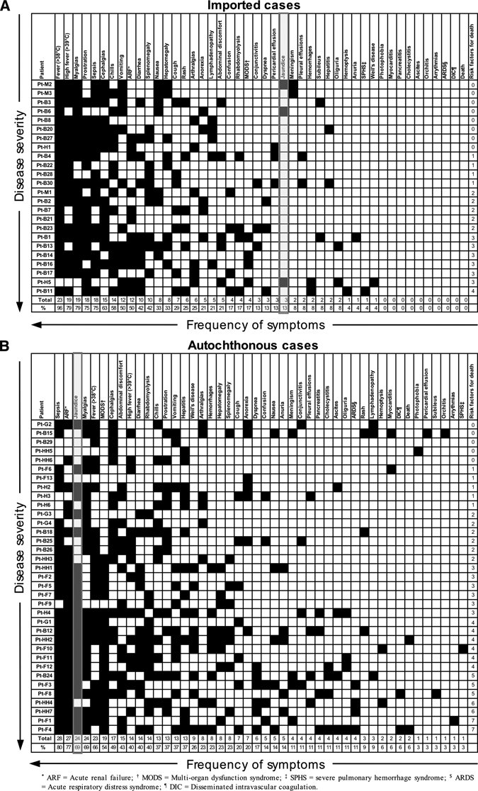
Disease severity (ordered by an increasing number of risk factors) and frequency of symptoms and syndromes associated with leptospirosis among patients with (A) imported and (B) autochthonous infections.
Statistics.
For boxplots, GraphPad PRISM (version 4.0, GraphPad Software, Inc., La Jolla, CA) and for statistical analysis, SPSS (version 16.0, SPSS Inc., Chicago, IL) were used. Fisher's exact test was applied for comparison of categorical variables and the Mann-Whitney U test for comparison of quantitative variables between the two subgroups. Two-tailed P values < 0.05 were considered statistically significant.
Results
Demographics.
In total, 59 adult patients, 8 women (14%) and 51 men (86%) between 18 and 71 years of age (median age 42 years) were included in this study (Figure 1, Table 2). Twenty-four patients (41%) contracted the disease outside (imported infections), 35 (59%) inside (autochthonous infections) their home country Austria or Germany, respectively (Figure 1). Distribution of age and sex, both being important determinants for disease severity, did not differ significantly between both subgroups, although there was a trend toward older age among patients with autochthonous infections (P = 0.0519). In both subgroups time from onset of symptoms to presentation was identical (median 5 days, range 1–22 days), and there was no statistically significant difference in time required for establishment of the diagnosis (Table 2). Highest incidence rates were recorded between August and November (Figure 1). The single most common exposure risk was recreation with exposure to fresh water (e.g., swimming, rafting, canoeing) being the most prevalent source of infection (Figure 1). Patients with imported leptospirosis most frequently traveled to The Caribbean (10; 42%), followed by Asia (9; 38%), Eastern Europe (3; 13%), Central America (1; 4%), and South America (1; 4%).
Figure 1.
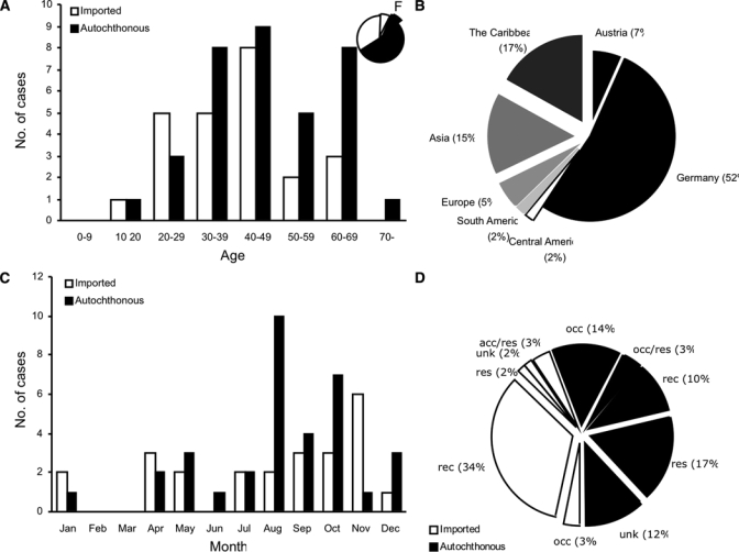
Demographics of patients with imported (N = 24) and autochthonous (N = 35) leptospirosis in Austria and Germany. (A) Distribution of age and sex. (B) Countries in which the diseases were contracted. (C) Seasonal distribution. (D) Mode of transmission. Acc = accidental transmission; acc/res = accidental and residential transmission probable; occ = occupational transmission; occ/res = occupational and residential transmission probable; res = residential transmission; unk = unknown.
Table 2.
Characteristics of the whole patient group, imported and autochthonous cases*
| Variable | Whole group (N = 59) | Imported cases (N = 24) | Autochthonous cases (N = 35) | Statistically significant differences between imported and autochthonous cases, P value |
|---|---|---|---|---|
| Demographics | ||||
| Age [years]: range (median) | 18–71 (42) | 19–62 (40) | 18–71 (47) | NS |
| Sex: F/M | 8/51 | 4/24 | 4/35 | NS |
| Time to presentation [days]: range (median) | 1–22 (5) | 2–18 (5) | 1–22 (5) | NS |
| Time to diagnosis [days]: range (median) | 1–39 (7) | 2–33 (8) | 1–39 (6) | NS |
| Risk factors | ||||
| Total number of independent risk factors for death: range (median) | 0–7 (2) | 0–4 (2) | 0–7 (3) | P = 0.0080 |
| WBC on admission [/nL]: range (median) | 2.80–20.60 (9.25) | 2.98–19.40 (9.00) | 2.80–20.60 (10.90) | NS |
| Altered mental status (%) | 9 (15) | 5 (21) | 4 (11) | NS |
| Hypotension/shock on admission (%) | 0 (0) | 0 (0) | 0 (0) | NS |
| Repolarization abnormalities on ECG (%) | 0 (0) | 0 (0) | 0 (0) | NS |
| Arrhythmias (%) | 1 (2) | 0 | 1 (3) | NS |
| Dyspnea (%) | 9 (15) | 3 (13) | 6 (17) | NS |
| Respiratory inufficiency (%) | 8 (14) | 1 (4) | 7 (20) | NS |
| K+ on admission [mmol/L]: range (median) | 2.60–4.70 (3.80) | 3.35–4.50 (3.90) | 2.60–4.70 (3.70) | NS |
| Oliguric ARF | 12 (20) | 3 (13) | 9 (26) | NS |
| Maximum serum creatinine level [mg/dL]: range (median) | 0.60–12.89 (2.62) | 0.60–10.31 (1.60) | 0.70–12.89 (4.85) | P = 0.0094 |
| BUN on admission [mg/dL]: range (median) | 13–355 (76) | 17–259 (60) | 13–355 (111) | P = 0.0057 |
| Disease severity | ||||
| Mild/asymptomatic (%) | 1 (2) | 0 (0) | 1 (3) | NS |
| Moderate/self-limited (%) | 12 (20) | 8 (33) | 4 (11) | NS |
| Severe/life-threatening (%) | 46 (78) | 16 (67) | 30 (86) | NS |
| Jaundice | ||||
| Jaundice (%): | 27 (46) | 3 (13) | 24 (69) | P = 0.0002 |
| Maximum serum bilirubine level [mg/dL]: range (median) | 0.4–69.1 (2.5) | 0.5–8.31 (1.15) | 0.4–69.1 (11.1) | P < 0.0001 |
| Outcome | ||||
| Length of hospital stay [days]: range (median) | 0-50 (11) | 0–36 (10) | 0–50 (12) | NS |
| Need of intensive care treatment | 24 (41) | 7 (29) | 17 (49) | NS |
| Length of stay on intensive care unit [days]: range (median) | 0–33 (0) | 0–14 (0) | 0–33 (0) | NS |
| Dialysis (%) | 12 (20) | 2 (8) | 10 (29) | NS |
| Need of mechanical ventilation (%) | 9 (15) | 1 (4) | 8 (23) | NS |
| Death (%) | 2 (3) | 0 (0) | 2 (6) | NS |
NS = statistically not significant; WBC = white blood cells; ECG = electrocardiogram; ARF = acute renal failure; BUN = blood urea nitrogen.
Symptoms and syndromes.
Figure 2 depicts typical leptospirosis-related symptoms and syndromes among patients with imported and autochthonous infections. In Figure 2 patients are ordered according to an increasing number of independent risk factors for death, symptoms, and syndromes according to their respective frequency. Fever (96%), myalgias (79%), typically located in the lumbar region and the calves, and prostration (75%) were the three most frequent symptoms among the 24 patients with imported infections, whereas sepsis (80%), ARF (77%), and jaundice (69%) were the most common clinical signs and syndromes in patients with autochthonous leptospirosis. Remarkably, only three of the 24 patients with imported leptospirosis (13%) became jaundiced, and all imported cases with severe/life-threatening disease remained anicteric, with the exception of a single patient with Weil's disease. Other gastrointestinal manifestations (nausea, vomiting, diarrhea, abdominal discomfort) were also more common in autochthonous compared with imported cases: in four cases each (11%) acute pancreatitis and cholecystitis with concomittant ascites occurred. Progression to Weil's disease occurred in nine icteric patients (26%) with autochthonous infections.
Impaired renal function was the most frequent single organ failure in both subgroups. Among imported infections, 12 patients (50%) had evidence of renal function impairment. Two patients (8%) developed oliguria, 1 (4%) anuria, 2 (8%) required dialysis. In autochthonous cases, renal function impairment was evident in 27 patients (77%), leading to oliguria in 5 (14%), and anuria in 4 patients (11%). Eighteen (75%) patients with imported infections versus 28 (80%) of autochthonous cases met the criteria of sepsis, 4 (17%) of imported versus 19 (54%) of autochthonous cases met the criteria of MODS either on admission or in the course of the illness. In two autochthonous cases (6%), Weil's disease was complicated by disseminated intravascular coagulation (DIC). An SPHS occurred in one case each of imported and autochthonous infection. Although some cases were hypotensive on admission, none of our patients required vasopressors to maintain a sufficient blood pressure. Cardiovascular complications were rare and only occurred within autochthonous cases: In two autochthonous patients (6%), myocarditis was diagnosed. One patient (3%) had ventricular tachycardia, whereas significant repolarization abnormalities were not documented in any case. Generally, the clinical spectrum appeared to be more divers in autochthonous versus imported disease (Figure 2).
Risk factors.
Forty-six (78%) of all patients were classified as cases with severe/life-threatening disease (i.e., presence of one or more independent risk factors for death): 16 (67%) of imported and 30 (86%) of autochthonous cases (no statistically significant difference; Table 2). With respect to laboratory parameters, patients with autochthonous infections had statistically significant higher creatinine and blood urea nitrogen (BUN) levels and lower platelet counts on admission and in the course of the illness (Figure 3). Both subgroups did not differ in a statistically significant way in the independent risk factors serum potassium and white blood cell (WBC) count on admission. In comparison to imported cases, patients with autochthonous infections had higher total numbers of independent risk factors for death (3 versus 2, P = 0.008; Table 2; Figure 4). Strikingly, jaundice was significantly more frequent among those who acquired the infection within Austria and Germany (24 of 35; 69%) than in those infected abroad (3 of 24; 13%; P < 0.0001). The possibility that bilirubin levels and jaundice might have been influenced by age was excluded be regression analysis (R2 = 0.001, P = 0.84). In multivariate analysis including age, proportions of icteric versus anicteric cases still differed significantly between autochthonous and imported cases (data not shown).
Figure 3.
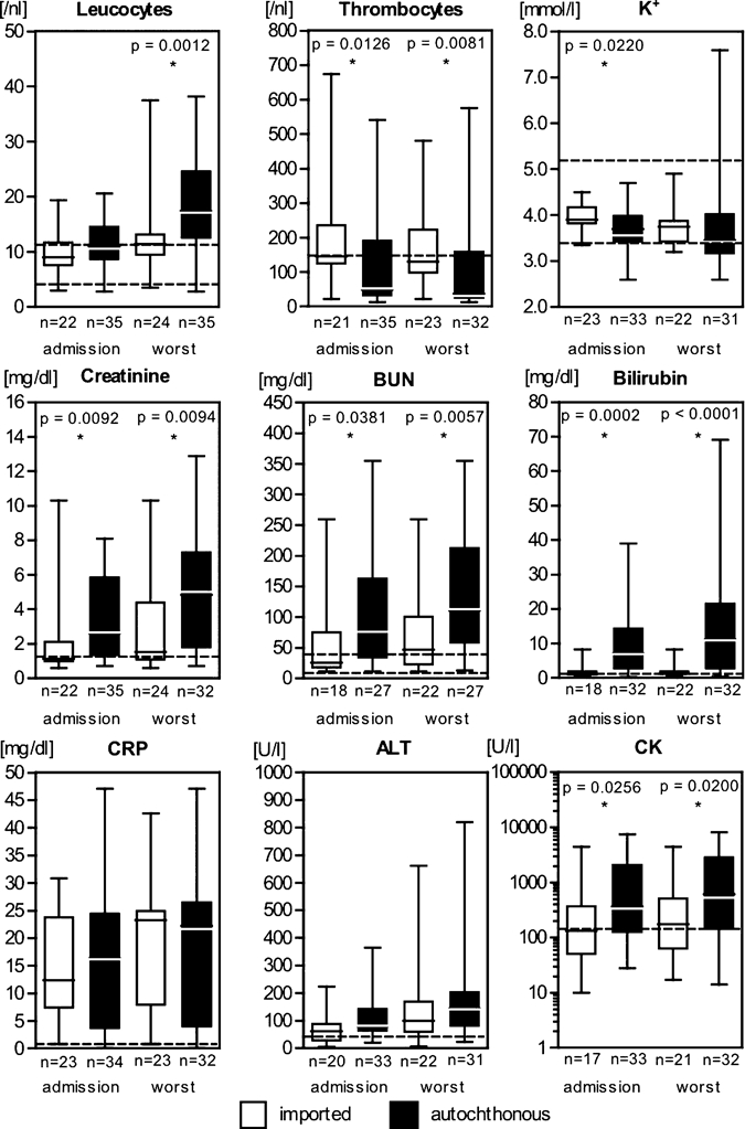
Relevant laboratory parameters of patients with imported (N = 24) and autochthonous (N = 35) leptospirosis in Austria and Germany. In each panel values on admission are represented by the left pair of boxplots, worst values in the course of the disease by the right pair of boxplots. Statistically significant differences are indicated by an asterix (*) with P values of Mann-Whitney U tests given. Dotted lines indicate reference values of conventional units. K+ = potassium; BUN = blood urea nitrogen; CRP = C-reactive protein; ALT = alaninaminotransferase; CK = creatinkinase.
Figure 4.
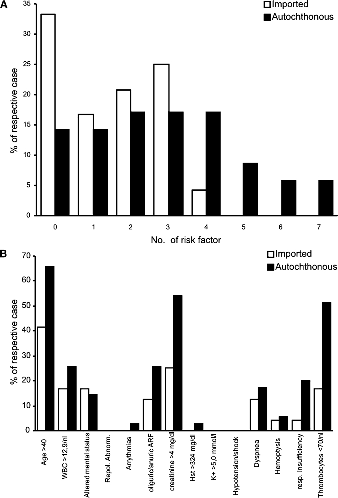
Total number of independent risk factors for (A) death and (B) frequency of individual risk factors among patients with imported (N = 24) and autochthonous leptospirosis (N = 35) in Austria and Germany. WBC = white blood cell count on admission; ARF = acute renal failure; BUN = blood urea nitrogen, K+ = potassium on admission.
Outcome.
Despite the fact that there were no significant differences between the subgroups with respect to overall disease severity according to pre-defined definitions, autochthonous cases tended to require longer hospitalization periods, longer intensive care treatment, and more frequent dialysis and mechanical ventilation (Table 2). There were no fatalities among patients with imported infections, whereas two patients with autochthonous infections (6%) died (Table 2): one because of cerebral mass bleeding, another due to septic shock with multi-organ failure.
Discussion
The aim of this study was to assess differences in clinical manifestations between imported and autochthonous cases of leptospirosis analyzing cases in Austria and Germany. The most important finding is that the cardinal clinical symptom of jaundice was significantly more frequent among autochthonous cases compared with imported infections.
Because of its unspecific and divers clinical presentation, leptospirosis is an under-recognized disease. Jaundice together with signs of a bacterial infection and only minor elevations in transaminase levels is not only considered as a cardinal symptom but also as an indicator of severe leptospirosis. Absence of jaundice therefore bears the potential of missing or delaying diagnosis and, hence, inadequate treatment. Two epidemics of leptospirosis showed the importance of jaundice as a clinical marker: In 1996, the high rate of jaundiced patients facilitated differentiation from those suffering from the parallel outbreak of dengue fever in Salvador, Brazil.17 In contrast, a high number of severely ill patients suffering from pulmonary hemorrhage without jaundice were described in an epidemic of leptospirosis in 1995 in Nicaragua.11,12 This clinical manifestation of leptospirosis has to be differentiated from other infections such as viral hemorrhagic fevers being a major concern for public health.
In the patients included in this study, demographics, seasonal trends, and exposure risks were consistent with the findings presented by Jansen and others27 for Germany. The majority of patients with autochthonous infections had severe forms with renal impairment, rhabdomyolysis, thrombocytopenia, Weil's disease, and serious pulmonary damage such as hemorrhagic pneumonitis, SPHS, and ARDS. Thirty patients had one or more risk factors for a fatal outcome of the disease, with two of them dying of Weil's disease. The majority of patients in this subgroup were icteric.
Patients with imported infections tended to present with less severe symptoms and mostly without jaundice. Still, there were life-threatening courses in this subgroup as well, with ARF, mental impairment, and SPHS. Examples are patient Pt-B1 with anuric ARF, rhabdomyolysis, thrombocytopenia, subileus, and pulmonary involvement after taking part in a marathon in the Philippine jungle, or patient Pt-B11 with severe respiratory insufficiency caused by SPHS and altered mental status. Similarly, some severely ill patients with autochthonous infections such as Pt-F12 and Pt-HH4 remained anicteric, whereas some patients with imported infections and only mild symptoms were jaundiced (e.g., Pt-B6). Jaundice could thus not clearly be assigned to severity of illness in either group. Notably, jaundice could not yet be identified as a marker of prognostic relevance, although this was repeatedly investigated.17,25
The exact reasons not only for the wide range of different courses of leptospirosis but also for the occurrence of jaundice are still unclear. The pathomechanisms of Leptospira interrogans are complex and not yet completely understood.3 The variability of the clinical course and severity of illness are most likely the result of a complex interplay between both bacterial and host factors. Only recently the genomes of various Leptospira species including L. interrogans serovar Lai have been sequenced completely, providing a genomic basis for more detailed analysis.33 Among the virulence factors identified so far are surface proteins, lipopolysaccharides, motility, chemotaxis, and secretory proteins such as sphingomyelinases, phospholipase C, and hemolysins including a pore-forming protein.34 The in vivo significance of any of these factors, however, has to be determined.3 An association of infecting serovar and disease severity has long been assumed, but could not be proven yet.8,18,35 In addition, host factors such as higher age17,23 and male sex36 predispose for more severe disease. In vitro, two more factors have been identified: serum levels of mannose-binding lectin,37 and toll-like receptor 4 (TLR4) linked activation of macrophages by whole heat-killed Leptospira as part of the innate immune system.38 The latter suggests that TLR4-receptors polymorphisms could add to the severity of leptospirosis.
There are two suspected main reasons for the occurrence of jaundice in leptospirosis: damage to liver architecture and, to a lesser extent, hemolysis, both potentially leading to a massive increase in serum bilirubin levels. Because of their mobility, Leptospira are able to invade liver sinuses and spaces of Dissé destroying the architecture of the canaliculi biliferi. Consequently, bilirubin passes into the blood.15 The bacteria can also invade hepatocytes, but a direct massive liver destruction usually does not occur, explaining the combination of massively increased bilirubin levels (in our study up to 70 mg/dL) with only moderately elevated liver enzymes (Figure 3). Leptospira can induce hepatocyte apoptosis,39 which could lead to a limitation in the local inflammatory response and, as a consequence, an increase of bacterial load in the liver. This local increase of bacterial load is assumed to add to systemic morbidity, which could explain the traditional concept of jaundice being a marker for the severity of the disease.15,39
Several studies have identified risk factors indicative of a severe course of leptospirosis, suggesting immediate transfer to intensive care of patients with those risk factors.18,20,21,26 We assessed disease severity by quantification of independent risk factors for death. A recent work by Ko and others17 suggests that certain combinations of risk factors may be more predictive of fatal outcome than others. Ko and others showed that oliguria was the strongest single predictor and that the presence of the three strongest predictors (oliguria, altered mental status, and age > 36 years) had an 82% positive predictive value for death. Because of the low death rate in our patients we could not account for this combinative effect. Further studies will be needed to determine the role of those combinations. A large proportion of our patients, however, presented with sepsis, in some complicated by multi-organ dysfunction, requiring immediate therapeutic interventions.
Treatment of leptospirosis mainly consists of aggressive symptomatic measures such as intensive care, fluid resuscitation, and correction of electrolyte disturbances, dialysis, and mechanical ventilation as needed. There is currently no evidence that specific antibiotic treatment improves outcome.40 In addition, there is general agreement that therapy is best initiated within 4 or less days after onset of symptoms to minimize immunologically mediated organ damage. The majority of patients in this study had a severe course of leptospirosis, but mortality rate was as low as 3%. We think that even without considering leptospirosis, these patients may have undergone optimal treatment as 1) risk factors identified for a possibly severe course of the disease are similar or even identical with the criteria of severe sepsis and MODS and 2) treatment of severe leptospirosis does not differ greatly from the general treatment strategy of sepsis. Both require intensive medical care, and commonly used regimes to treat community-acquired sepsis with unknown pathogens (including aminopenicillines with betalactamase-inhibitors or third generation cephalosporins) will cover the bacteria as well.
Data presented here are the result of a retrospective study with attendant limitations. The approach of a multi-centered analysis in two countries should in part limit biased case selection. As the participating hospitals are tertiary care centers, there might be a bias toward more severe cases. The study sample includes about 10% of all cases notified in Austria and Germany between 1998 and 2008. However, all limitations related to retrospective analyses apply.
In conclusion, leptospirosis in Austria and Germany is an emerging infectious disease, both imported and autochthonous in origin, variable in its presentation with a significant and presumably increasing proportion of severe anicteric cases.
Acknowledgments
We thank Joel B. Lande for his help in preparing the manuscript.
Footnotes
Authors' addresses: Bodo Hoffmeister and Norbert Suttorp, Department of Infectious Diseases and Pulmonology, Charité University Medicine Berlin, Berlin, Germany, E-mails: b.hoffmeister-infekt@charite.de and norbert.suttorp@charite.de. Gabriele Peyerl-Hoffmann and Winfried V. Kern, Center for Infectious Diseases and Travel Medicine, Department of Medicine, University Hospital Freiburg, Freiburg, Germany, E-mails: gabriele.peyerl-hoffmann@uniklinik-freiburg.de and kern@if-freiburg.de. Sven Pischke, Department of Gastroenterology, Hepatology and Endocrinology, Hannover Medical School, Hannover, Germany, E-mail: pischke.sven@mh-hannover.de. Ines Zollner-Schwetz and Robert Krause, Section of Infectious Diseases, Division of Pulmonology, Department of Internal Medicine, Medical University of Graz, Graz, Austria, E-mails: ines.schwetz@meduni-graz.at and robert.krause@medunigraz.at. Matthias C. Müller, Department of Infectious Diseases and Tropical Medicine, Ludwig-Maximilians University of Munich, Munich and Department of Infectious Diseases, Medizinische Poliklinik, Ludwig-Maximilians University of Munich, Munich, Germany, E-mail: matthias.mueller@med.uni-muenchen.de. Angelika Graf, Department of Anaesthesiology and Intensive Care Medicine, Charité University Medicine Berlin, Berlin, Germany, E-mail: angelika.graf@charite.de. Stefan Kluge, Department of Intensive Care, University Medical Center Hamburg-Eppendorf, Hamburg, Germany, E-mail: s.kluge@uke.de. Gerd D. Burchard and Jakob P. Cramer, Section Tropical Medicine, Department of Internal Medicine, University Medical Center Hamburg-Eppendorf, Hamburg, Germany and Section Tropical Medicine/Clinical Research, Bernhard-Nocht-Institute for Tropical Medicine, Hamburg, Germany, E-mails: gerd.burchard@bni-hamburg.de and cramer@bni-hamburg.de.
Reprint requests: Bodo Hoffmeister, Department of Infectious Diseases and Pulmonology, Charité University Medicine Berlin, Augustenburger Platz 1, 13353 Berlin, Germany, Tel: +49-30-450653389, Fax: +49-30-450553958, E-mail: b.hoffmeister-infekt@charite.de.
References
- 1.1999. Leptospirosis worldwide Wkly Epidemiol Rec. 1999;74:237–242. [PubMed] [Google Scholar]
- 2.Levett PN. Leptospirosis. Clin Microbiol Rev. 2001;14:296–326. doi: 10.1128/CMR.14.2.296-326.2001. [DOI] [PMC free article] [PubMed] [Google Scholar]
- 3.Bharti AR, Nally JE, Ricaldi JN, Matthias MA, Diaz MM, Lovett MA, Levett PN, Gilman RH, Willig MR, Gotuzzo E, Vinetz JM. Leptospirosis: a zoonotic disease of global importance. Lancet Infect Dis. 2003;3:757–771. doi: 10.1016/s1473-3099(03)00830-2. [DOI] [PubMed] [Google Scholar]
- 4.Peter G. Leptospirosis: a zoonosis of protean manifestations. Pediatr Infect Dis. 1982;1:282–288. [PubMed] [Google Scholar]
- 5.Turner LH. Leptospirosis. I. Trans R Soc Trop Med Hyg. 1967;61:842–855. doi: 10.1016/0035-9203(67)90045-4. [DOI] [PubMed] [Google Scholar]
- 6.Bovet P, Yersin C, Merien F, Davis CE, Perolat P. Factors associated with clinical leptospirosis: a population-based case-control study in the Seychelles (Indian Ocean) Int J Epidemiol. 1999;28:583–590. doi: 10.1093/ije/28.3.583. [DOI] [PubMed] [Google Scholar]
- 7.Ashford DA, Kaiser RM, Spiegel RA, Perkins BA, Weyant RS, Bragg SL, Plikaytis B, Jarquin C, De Lose Reyes JO, Amador JJ. Asymptomatic infection and risk factors for leptospirosis in Nicaragua. Am J Trop Med Hyg. 2000;63:249–254. [PubMed] [Google Scholar]
- 8.Paganin F, Bourdin A, Dalban C, Courtin JP, Poubeau P, Borgherini G, Michault A, Sally JC, Tixier F, Genin R, Arvin-Berod C. Leptospirosis in Reunion Island (Indian Ocean): analysis of factors associated with severity in 147 confirmed cases. Intensive Care Med. 2007;33:1959–1966. doi: 10.1007/s00134-007-0776-y. [DOI] [PubMed] [Google Scholar]
- 9.Wang CN, John L, Chang TF, Cheng WJ, Lou MY, Hung AT. Studies on anicteric leptospirosis. I. Clinical manifestations and antibiotic therapy. Chin Med J (Engl) 1965;84:283–291. [PubMed] [Google Scholar]
- 10.Park SK, Lee SH, Rhee YK, Kang SK, Kim KJ, Kim MC, Kim KW, Chang WH. Leptospirosis in Chonbuk Province of Korea in 1987: a study of 93 patients. Am J Trop Med Hyg. 1989;41:345–351. [PubMed] [Google Scholar]
- 11.Zaki SR, Shieh WJ. Leptospirosis associated with outbreak of acute febrile illness and pulmonary haemorrhage, Nicaragua, 1995. The Epidemic Working Group at Ministry of Health in Nicaragua. Lancet. 1996;347:535–536. doi: 10.1016/s0140-6736(96)91167-8. [DOI] [PubMed] [Google Scholar]
- 12.Trevejo RT, Rigau-Perez JG, Ashford DA, McClure EM, Jarquin-Gonzalez C, Amador JJ, de los Reyes JO, Gonzalez A, Zaki SR, Shieh WJ, McLean RG, Nasci RS, Weyant RS, Bolin CA, Bragg SL, Perkins BA, Spiegel RA. Epidemic leptospirosis associated with pulmonary hemorrhage-Nicaragua, 1995. J Infect Dis. 1998;178:1457–1463. doi: 10.1086/314424. [DOI] [PubMed] [Google Scholar]
- 13.Segura ER, Ganoza CA, Campos K, Ricaldi JN, Torres S, Silva H, Cespedes MJ, Matthias MA, Swancutt MA, Lopez Linan R, Gotuzzo E, Guerra H, Gilman RH, Vinetz JM. Clinical spectrum of pulmonary involvement in leptospirosis in a region of endemicity, with quantification of leptospiral burden. Clin Infect Dis. 2005;40:343–351. doi: 10.1086/427110. [DOI] [PMC free article] [PubMed] [Google Scholar]
- 14.Kuriakose M, Eapen CK, Paul R. Leptospirosis in Kolenchery, Kerala, India: epidemiology, prevalent local serogroups and serovars and a new serovar. Eur J Epidemiol. 1997;13:691–697. doi: 10.1023/a:1007300729615. [DOI] [PubMed] [Google Scholar]
- 15.Plank R, Dean D. Overview of the epidemiology, microbiology, and pathogenesis of Leptospira spp. in humans. Microbes Infect. 2000;2:1265–1276. doi: 10.1016/s1286-4579(00)01280-6. [DOI] [PubMed] [Google Scholar]
- 16.Daher E, Zanetta DM, Cavalcante MB, Abdulkader RC. Risk factors for death and changing patterns in leptospirosis acute renal failure. Am J Trop Med Hyg. 1999;61:630–634. doi: 10.4269/ajtmh.1999.61.630. [DOI] [PubMed] [Google Scholar]
- 17.Ko AI, Galvao Reis M, Ribeiro Dourado CM, Johnson WD, Jr, Riley LW. Urban epidemic of severe leptospirosis in Brazil. Salvador Leptospirosis Study Group. Lancet. 1999;354:820–825. doi: 10.1016/s0140-6736(99)80012-9. [DOI] [PubMed] [Google Scholar]
- 18.Dupont H, Dupont-Perdrizet D, Perie JL, Zehner-Hansen S, Jarrige B, Daijardin JB. Leptospirosis: prognostic factors associated with mortality. Clin Infect Dis. 1997;25:720–724. doi: 10.1086/513767. [DOI] [PubMed] [Google Scholar]
- 19.Panaphut T, Domrongkitchaiporn S, Thinkamrop B. Prognostic factors of death in leptospirosis: a prospective cohort study in Khon Kaen, Thailand. Int J Infect Dis. 2002;6:52–59. doi: 10.1016/s1201-9712(02)90137-2. [DOI] [PubMed] [Google Scholar]
- 20.Doudier B, Garcia S, Quennee V, Jarno P, Brouqui P. Prognostic factors associated with severe leptospirosis. Clin Microbiol Infect. 2006;12:299–300. doi: 10.1111/j.1469-0691.2005.01335.x. [DOI] [PubMed] [Google Scholar]
- 21.Marotto PC, Nascimento CM, Eluf-Neto J, Marotto MS, Andrade L, Sztajnbok J, Seguro AC. Acute lung injury in leptospirosis: clinical and laboratory features, outcome, and factors associated with mortality. Clin Infect Dis. 1999;29:1561–1563. doi: 10.1086/313501. [DOI] [PubMed] [Google Scholar]
- 22.Lopes AA, Costa E, Costa YA, Bina JC, Sacramento E. The association between serum potassium at hospital admission and the case-fatality rate of leptospirosis in men. Rev Inst Med Trop Sao Paulo. 2001;43:217–220. doi: 10.1590/s0036-46652001000400008. [DOI] [PubMed] [Google Scholar]
- 23.Spichler AS, Vilaca PJ, Athanazio DA, Albuquerque JO, Buzzar M, Castro B, Seguro A, Vinetz JM. Predictors of lethality in severe leptospirosis in urban Brazil. Am J Trop Med Hyg. 2008;79:911–914. [PMC free article] [PubMed] [Google Scholar]
- 24.Pappachan MJ, Mathew S, Aravindan KP, Khader A, Bharghavan PV, Kareem MM, Tuteja U, Shukla J, Batra HV. Risk factors for mortality in patients with leptospirosis during an epidemic in northern Kerala. Natl Med J India. 2004;17:240–242. [PubMed] [Google Scholar]
- 25.Tantitanawat S, Tanjatham S. Prognostic factors associated with severe leptospirosis. J Med Assoc Thai. 2003;86:925–931. [PubMed] [Google Scholar]
- 26.Esen S, Sunbul M, Leblebicioglu H, Eroglu C, Turan D. Impact of clinical and laboratory findings on prognosis in leptospirosis. Swiss Med Wkly. 2004;134:347–352. doi: 10.4414/smw.2004.10436. [DOI] [PubMed] [Google Scholar]
- 27.Jansen A, Schoneberg I, Frank C, Alpers K, Schneider T, Stark K. Leptospirosis in Germany, 1962–2003. Emerg Infect Dis. 2005;11:1048–1054. doi: 10.3201/eid1107.041172. [DOI] [PMC free article] [PubMed] [Google Scholar]
- 28.van Crevel R, Speelman P, Gravekamp C, Terpstra WJ. Leptospirosis in travelers. Clin Infect Dis. 1994;19:132–134. doi: 10.1093/clinids/19.1.132. [DOI] [PubMed] [Google Scholar]
- 29.Benzler J, Krause G. On the 2004 edition of case definitions for the surveillance of notifiable infectious diseases in Germany. Bundesgesundheitsblatt Gesundheitsforschung Gesundheitsschutz. 2004;47:141–146. doi: 10.1007/s00103-003-0783-2. [DOI] [PubMed] [Google Scholar]
- 30.Bellomo R, Ronco C, Kellum JA, Mehta RL, Palevsky P. Acute renal—definition, outcome measures, animal models, fluid therapy and information technology needs: the Second International Consensus Conference of the Acute Dialysis Quality Initiative (ADQI) Group. Crit Care. 2004;8:R204–R212. doi: 10.1186/cc2872. [DOI] [PMC free article] [PubMed] [Google Scholar]
- 31.Levy MM, Fink MP, Marshall JC, Abraham E, Angus D, Cook D, Cohen J, Opal SM, Vincent JL, Ramsay G. 2001 SCCM/ESICM/ACCP/ATS/SIS International Sepsis Definitions Conference. Crit Care Med. 2003;31:1250–1256. doi: 10.1097/01.CCM.0000050454.01978.3B. [DOI] [PubMed] [Google Scholar]
- 32.Bernard GR, Artigas A, Brigham KL, Carlet J, Falke K, Hudson L, Lamy M, Legall JR, Morris A, Spragg R. The American-European Consensus Conference on ARDS. Definitions, mechanisms, relevant outcomes, and clinical trial coordination. Am J Respir Crit Care Med. 1994;149:818–824. doi: 10.1164/ajrccm.149.3.7509706. [DOI] [PubMed] [Google Scholar]
- 33.Ren SX, Fu G, Jiang XG, Zeng R, Miao YG, Xu H, Zhang YX, Xiong H, Lu G, Lu LF, Jiang HQ, Jia J, Tu YF, Jiang JX, Gu WY, Zhang YQ, Cai Z, Sheng HH, Yin HF, Zhang Y, Zhu GF, Wan M, Huang HL, Qian Z, Wang SY, Ma W, Yao ZJ, Shen Y, Qiang BQ, Xia QC, Guo XK, Danchin A, Saint Girons I, Somerville RL, Wen YM, Shi MH, Chen Z, Xu JG, Zhao GP. Unique physiological and pathogenic features of Leptospira interrogans revealed by whole-genome sequencing. Nature. 2003;422:888–893. doi: 10.1038/nature01597. [DOI] [PubMed] [Google Scholar]
- 34.Ko AI, Goarant C, Picardeau M. Leptospira: the dawn of the molecular genetics era for an emerging zoonotic pathogen. Nat Rev Microbiol. 2009;7:736–747. doi: 10.1038/nrmicro2208. [DOI] [PMC free article] [PubMed] [Google Scholar]
- 35.Katz AR, Ansdell VE, Effler PV, Middleton CR, Sasaki DM. Assessment of the clinical presentation and treatment of 353 cases of laboratory-confirmed leptospirosis in Hawaii, 1974–1998. Clin Infect Dis. 2001;33:1834–1841. doi: 10.1086/324084. [DOI] [PubMed] [Google Scholar]
- 36.Jansen A, Stark K, Schneider T, Schoneberg I. Sex differences in clinical leptospirosis in Germany: 1997–2005. Clin Infect Dis. 2007;44:e69–e72. doi: 10.1086/513431. [DOI] [PubMed] [Google Scholar]
- 37.Miranda KA, Vasconcelos LR, Coelho LC, Lima Filho JL, Cavalcanti MS, Moura P. High levels of serum mannose-binding lectin are associated with the severity of clinical signs of leptospirosis. Braz J Med Biol Res. 2009;42:353–357. doi: 10.1590/s0100-879x2009000400007. [DOI] [PubMed] [Google Scholar]
- 38.Viriyakosol S, Matthias MA, Swancutt MA, Kirkland TN, Vinetz JM. Toll-like receptor 4 protects against lethal Leptospira interrogans serovar icterohaemorrhagiae infection and contributes to in vivo control of leptospiral burden. Infect Immun. 2006;74:887–895. doi: 10.1128/IAI.74.2.887-895.2006. [DOI] [PMC free article] [PubMed] [Google Scholar]
- 39.Merien F, Truccolo J, Rougier Y, Baranton G, Perolat P. In vivo apoptosis of hepatocytes in guinea pigs infected with Leptospira interrogans serovar icterohaemorrhagiae. FEMS Microbiol Lett. 1998;169:95–102. doi: 10.1111/j.1574-6968.1998.tb13304.x. [DOI] [PubMed] [Google Scholar]
- 40.Guidugli F, Castro AA, Atallah AN. Antibiotics for treating leptospirosis. Cochrane Database Syst Rev. 2000:CD001306. doi: 10.1002/14651858.CD001306. [DOI] [PubMed] [Google Scholar]