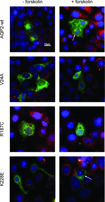
Figure 4. Immunofluorescence analysis of AQP2 in transfected IMCD-3 cells.

mIMCD-3 cells were transfected with pcDNA6-AQP2-wt, -V24A, -R187C or -K228E, incubated for 16–24 h and treated (+forskolin) or not (–forskolin) with forskolin (50 μm, 45 min) prior to fixation. PDI was used as an ER marker (red) and DAPI was used as a nuclear stain (blue). Plasma membrane staining is found for both AQP2-wt and -K228E (arrows) but not for R187C or V24A. Magnification ×60. Scale bar, 10 μm.
