
Figure 3. PAR agonists stimulate mucin release from Calu-3 cells.
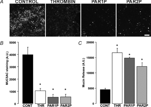
Calu-3 cells were challenged with vehicle, 50 nm thrombin, 100 μm PAR1P, or 100 μm PAR2P for 5 min at 37°C. A, mucin granule content was determined by immunostaining with a MUC5AC antibody followed by confocal microscopy analysis (bar = 100 μm). B, quantification of MUC5AC immunostaining in Calu-3 cultures. C, mucin release in the lumenal bath was assessed by slot blot as in Fig. 1. The results of a representative experiment are illustrated, and the data are expressed in arbitrary units and are the mean ±s.e.m. (n= 4; *P < 0.01). Similar results were obtained in three independent experiments.