
Figure 4. PAR-stimulated mucin release is Ca2+ and cytoskeleton dependent.
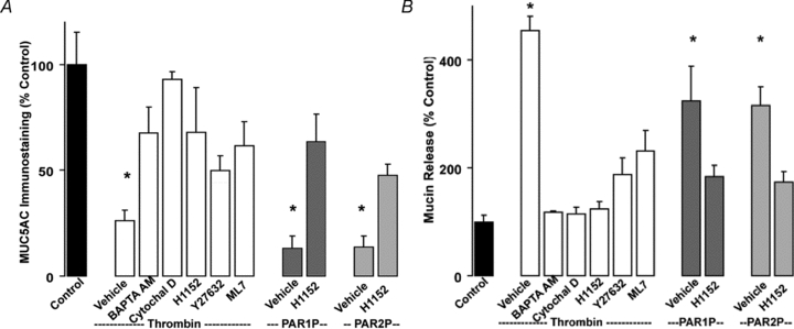
Calu-3 cells were pre-incubated for 30 min at 37°C with vehicle, 10 μm BAPTA AM, 5 μm cytochalasin D, 100 nm H1152, 10 μm Y27632, or 1 μm ML7. Cells were challenged with vehicle, 50 nm thrombin, 100 μm PAR1P, or 100 μm PAR2P for 5 min at 37°C. A, mucin granule content was quantified by immunostaining as in Fig. 3. B, mucin release in the apical bath was assessed by slot blot as in Fig. 1. Experiments were performed three times, each condition in quadruplicate. The results of a representative experiment are illustrated and data are expressed as percentage of control (mean ±s.e.m.; *P < 0.01 vs. control).