
Figure 5. PAR-stimulated ATP release involves a vesicular, Ca2+- and cytoskeleton- dependent mechanism.
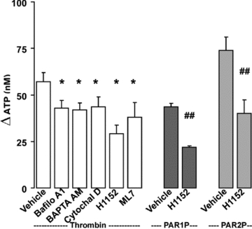
Calu-3 cells were pre-incubated with inhibitors as in Fig. 4, or with 4 μm Bafilomycin A1 for 30 min at 37°C. Mucosal ATP release following the addition of the indicated PAR agonists (5 min at 37°C) was assessed using the luciferin–luciferase assay in the presence of blockers of ecto-nucleotidases. Experiments were performed in quadruplicate with three independent cultures. The results of a representative experiment are illustrated, and the data are expressed as the difference between PAR agonist and basal values (basal ATP values, 15 ± 5 nm) (mean ±s.e.m.; *P < 0.01 compared to thrombin stimulation; ##P < 0.01 compared to PAR1P and PAR2P stimulation).