
FIG. 4.
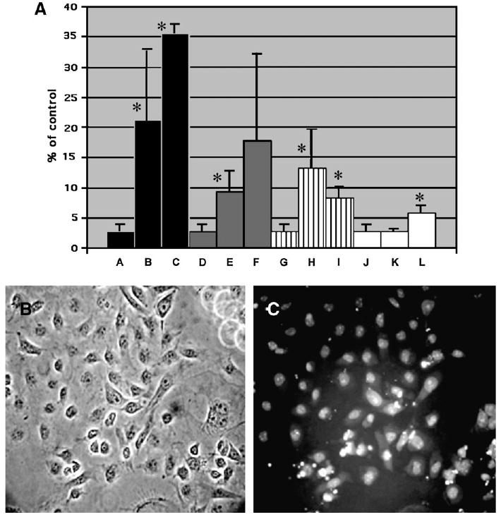
Induction of apoptosis by cadmium oxide and different types of nanoparticles in the C18–4 spermatogonial stem cells after 48 h incubation. A. At the end of the incubation period, the percentage of cells in apoptosis was measured with the Vybrant assay that uses the YO-PRO-1 dye and propidium iodide as described in Materials and Methods. The graph is a composite of two experiments, with n ≥ 3 for each point.*Statistically significant difference compared with controls (p < 0.05). Lanes: A. control (medium +PBS); B. cadmium oxide (1 μg/ml); C. cadmium oxide (5 μg/ml); D. control (medium + PBS); E. silver nanoparticles (5 μg/ml); F. silver nanoparticles (10 μg/ml); G. control (medium + PBS); H. aluminum nanoparticles (5 μg/ml); I. aluminum nanoparticles (10 μg/ml); J. control (medium + PBS); K. molybdenum nanoparticles (25 μg/ml). L. molybdenum nanoparticles (50 μg/ml). B. Morphology of the C18–4 spermatogonial stem cells in phase contrast microscopy after incubation with 10 μg/ml molybdenum nanoparticles for 48 h. (400×). C. Same field in fluoresence microscopy showing cells positive for the YO-PRO-1 dye (apoptotic cells). These cells are negative for propidium iodide.