Figure 1.
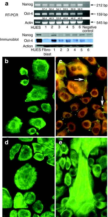
Characterization of primary human umbilical cord–lining epithelial cells (CLECs). (a) RT-PCR and western blot analysis of different CLEC samples (1–6), human embryonic stem cell line (HUES, positive control), and human primary dermal fibroblasts (negative control) for expression of Oct-4 and Nanog. Negative control for RT-PCR was a minus template PCR. Shown below RT-PCR gel images are quantitative levels of Oct-4 and Nanog transcripts (normalized to actin) relative to the HUES sample. Indirect immunofluorescence staining for (b) universal keratins; (c) desmoplakin (positive expression is seen as bright green fluorescence (arrows) and negative expression as dull orange); (d) keratin 18 and (e) keratin 19 in cultured CLECs. Original magnification ×400. bp, base pair.

