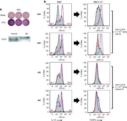
Figure 4.
Expansion of CD4+IL-10+FoxP3+ T-cells in response to canine FIX (cFIX) antigens in dogs receiving an AAV-2-cFIX vector via ATVRX. (a) Top: ELISpot assay for IL-10 secretion after stimulation with purified canine FIX protein. Bottom: western blot analysis of cFIX protein in liver lysates from normal and HB dogs. (b) Staining for CD4+IL-10+FoxP3+ T-cells after in vitro restimulation with either cFIX protein (red line), peptide 68 from cFIX (blue line), or cFIX irrelevant pool (gray shaded area). Left panels, histogram plots showing IL-10 intracellular staining on CD4+ T-cells; right panels, FoxP3 staining on cells gated on CD4+IL-10+ T-cells. Cells were gated on lymphocytes, CD4+CD8−. ATVRX, afferent transvenular retrograde extravasation; AAV, adeno-associated virus; ELISpot, enzyme-linked immunosorbent spot; HB, hemophilia B; IL, interleukin.

