FIGURE 2.
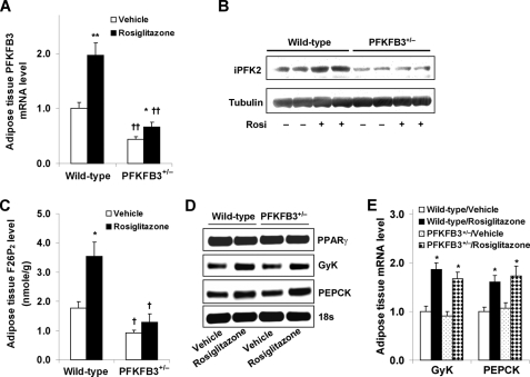
Disruption of PFKFB3/iPFK2 impairs the response of adipose tissue PFKFB3/iPFK2 but not other PPARγ target genes to PPARγ activation. At the age of 5–6 weeks, male PFKFB3+/− mice and wild-type littermates were fed an HFD for 12 weeks and treated with rosiglitazone (10 mg/kg/day) or vehicle (PBS) during the last 4 weeks of HFD feeding. At the end of the feeding/treatment regimen, mice were fasted for 4 h before collection of tissue samples. Epididymal adipose tissue samples were used for the analyses. A, the mRNA levels of PFKFB3 were measured using real-time RT-PCR. B, adipose tissue iPFK2 was determined using Western blot. C, adipose tissue F26P2 levels were determined using the 6-phosphofructo-1-kinase activation method. D, representative PCR products of adipose tissue genes. E, quantification of the expression of adipose tissue genes. Rosi, rosiglitazone. For A, C, and E, data are means ± S.E. (error bars), n = 6. *, p < 0.05; **, p < 0.01, rosiglitazone versus vehicle within the same genotype (in A and C) or wild type/rosiglitazone versus wild type/vehicle and PFKFB3+/−/rosiglitazone versus PFKFB3+/−/vehicle for the same gene (in E). †, p < 0.05; ††, p < 0.01, PFKFB3+/− versus wild type with the same treatment (rosiglitazone or vehicle in A and C).

