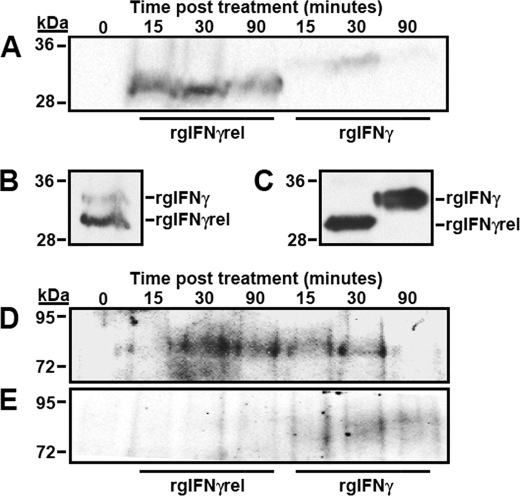
FIGURE 6.
Analysis of rgIFNγrel and rgIFNγ cellular association, Stat1 tyrosine phosphorylation and phospho-(Y)-Stat1 nuclear accumulation in monocytes treated with rgIFNγrel or rgIFNγ. Five million monocytes were incubated with either medium alone, 5 μg of rgIFNγrel or 5 μg of rgIFNγ for 0, 15, 30, or 90 min. Whole cell lysates (A) were assayed by Western blot using α-polyHis antibody. Cells were also co-incubated with 5 μg of rgIFNγrel and 5 μg of rgIFNγ for a half-hour (B). The relative amounts of rgIFNγrel and rgIFNγ added to cells can be seen in C. Five million monocytes were incubated with either medium, 100 ng/ml of rgIFNγrel, or 100 ng/ml of rgIFNγ for 0, 15, 30, or 90 min. Whole cell lysates (D) or isolated nuclei (E) were assessed by Western blot with an α-phospho-(Tyr)-Stat1 antibody.

