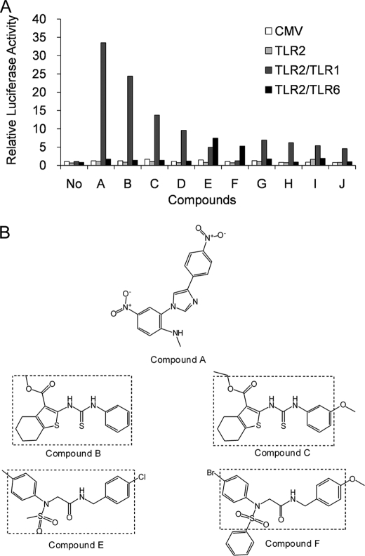
FIGURE 1.
Activity and structure of novel chemical agonists for TLR2. A, SW620 cells were co-transfected with various combinations of TLRs and an IL-8-driven luciferase gene and seeded into a 384-well plate. Two days after transfection, cells were stimulated with the indicated compounds at a concentration of ∼100 μm for at least 6 h. Cells were then lysed, and luciferase activities were measured. All values were normalized to those of unstimulated cells with reporter and empty FLAG-CMV vector (CMV). Each bar represents the average of two independent experimental values. B, the chemical structures of the novel TLR2 agonists. Dashed lines show the common structures identified for certain pairs of compounds.

