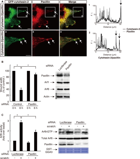
FIGURE 5.
Cytohesin-2, acting through Arf6, cooperates with paxillin at the leading edge of migrating 3T3-L1 cells. A, the plasmid encoding GFP-tagged cytohesin-2 was electroporated into 3T3-L1 cells, and cells were immunostained using anti-GFP antibody and anti-paxillin antibody 6 h after scratching. Representative confocal images of GFP-tagged cytohesin-2 (green) and endogenous paxillin (red) were collected with a confocal microscope system. Staining intensities measured according to pixel brightness were evaluated through a fluorescence intensity (F.I.) profile of a typical line scan (lines 1 and 2). The lower panels (a) are high magnification images of the areas boxed with white lines in the corresponding upper panels. The scale bar indicates 50 μm. B and C, 3T3-L1 cells were transfected with an siRNA for control (luciferase) or paxillin. B, cell monolayers were wounded by scraping and observed microscopically at 0 and 6 h. The rate of healing was calculated based on the width of the wound. C, after 0 or 30 scratches, the Arf6 GTP form was measured with GST-GGA3, respectively. Cell lysates were immunoblotted with an antibody against paxillin, small GTP-binding protein, or actin. Data were evaluated with one-way ANOVA (*, p < 0.01).

